[English] 日本語
Yorodumi
- PDB-2wkc: Crystal structure from a single-stranded DNA binding protein from... -

+
Open data


ID or keywords:

Loading...

-
Basic information

Entry
Database: PDB / ID: 2wkc
Title
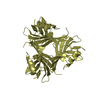
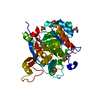
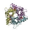
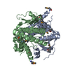
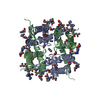
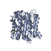
Crystal structure from a single-stranded DNA binding protein from the lactococcal phage p2
ComponentsORF34P2
KeywordsDNA BINDING PROTEIN / SINGLE-STRANDED DNA BINDING / SSB / LACTOCOCCAL PHAGE PROTEIN
Function / homologyLactococcus phage single-stranded DNA binding protein / Lactococcus phage single-stranded DNA binding protein / Lactococcus phage SSB superfamily / Lactococcus phage single-stranded DNA binding protein / OB fold (Dihydrolipoamide Acetyltransferase, E2P) / Beta Barrel / Mainly Beta / SSB protein
Function and homology information
Biological speciesLACTOCOCCUS PHAGE P2 (virus)
MethodX-RAY DIFFRACTION / SYNCHROTRON / MOLECULAR REPLACEMENT / Resolution: 2.6 Å
AuthorsScaltriti, E. / Cambillau, C. / Ortiz-Lombardia, M.
CitationJournal: Mol.Microbiol. / Year: 2009
Title: Structure and Function of Phage P2 Orf34(P2), a New Type of Single-Stranded DNA Binding Protein.
Authors: Scaltriti, E. / Tegoni, M. / Rivetti, C. / Launay, H. / Masson, J.Y. / Magadan, A.H. / Tremblay, D. / Moineau, S. / Ramoni, R. / Lichiere, J. / Campanacci, V. / Cambillau, C. / Ortiz-Lombardia, M.
History
DepositionJun 9, 2009Deposition site: PDBE / Processing site: PDBE
Revision 1.0Sep 15, 2009Provider: repository / Type: Initial release
Revision 1.1Jul 13, 2011Group: Advisory / Version format compliance
Revision 1.2May 29, 2019Group: Data collection / Experimental preparation / Other
Category: exptl_crystal_grow / pdbx_database_proc ...exptl_crystal_grow / pdbx_database_proc / pdbx_database_status / struct_biol
Item: _exptl_crystal_grow.method / _pdbx_database_status.recvd_author_approval
Revision 1.3Dec 13, 2023Group: Data collection / Database references ...Data collection / Database references / Other / Refinement description
Category: chem_comp_atom / chem_comp_bond ...chem_comp_atom / chem_comp_bond / database_2 / pdbx_database_status / pdbx_initial_refinement_model
Item: _database_2.pdbx_DOI / _database_2.pdbx_database_accession / _pdbx_database_status.status_code_sf
Remark 700 SHEET THE SHEET STRUCTURE OF THIS MOLECULE IS BIFURCATED. IN ORDER TO REPRESENT THIS FEATURE IN ... SHEET THE SHEET STRUCTURE OF THIS MOLECULE IS BIFURCATED. IN ORDER TO REPRESENT THIS FEATURE IN THE SHEET RECORDS BELOW, TWO SHEETS ARE DEFINED.

-
Structure visualization

Structure viewerMolecule:
MolmilJmol/JSmol

Downloads & links

-
Assembly

Deposited unit
A: ORF34P2
B: ORF34P2
C: ORF34P2
D: ORF34P2


Theoretical massNumber of molelcules
Total (without water)51,7494
Polymers51,7494
Non-polymers00
Water50428
1


  • Idetical with deposited unit
  • defined by author&software
TypeNameSymmetry operationNumber
identity operation1_555x,y,z1
Buried area7720 Å2
ΔGint-26.77 kcal/mol
Surface area19750 Å2
MethodPISA
Unit cell
Length a, b, c (Å)48.280, 71.710, 109.730
Angle α, β, γ (deg.)90.00, 90.00, 90.00
Int Tables number19
Space group name H-MP212121

-
Components

#1: Protein
ORF34P2


Mass: 12937.332 Da / Num. of mol.: 4 / Fragment: RESIDUES 15-131
Source method: isolated from a genetically manipulated source
Source: (gene. exp.) LACTOCOCCUS PHAGE P2 (virus) / Plasmid: PETG-20A / Production host: ESCHERICHIA COLI (E. coli) / Strain (production host): ROSETTA (DE3) PLYSS / References: UniProt: Q09WL7
#2: Water ChemComp-HOH / water


Mass: 18.015 Da / Num. of mol.: 28 / Source method: isolated from a natural source / Formula: H2O

-
Experimental details

-
Experiment

ExperimentMethod: X-RAY DIFFRACTION / Number of used crystals: 1

-
Sample preparation

CrystalDensity Matthews: 2.76 Å3/Da / Density % sol: 55.5 % / Description: NONE
Crystal growMethod: vapor diffusion, sitting drop / pH: 6.2
Details: PROTEIN AT 7 MG/ML IN 10 MM TRIS, 300 MM NACL PH 8.0 WAS CRYSTALLIZED BY SITTING-DROP VAPOUR DIFFUSION AGAINST 0.1 M SODIUM CACODYLATE PH 6.2, 45% MME-PEG2000

-
Data collection

DiffractionMean temperature: 100 K
Diffraction sourceSource: SYNCHROTRON / Site: ESRF / Beamline: ID23-2 / Wavelength: 0.8726
DetectorType: MARMOSAIC 225 mm CCD / Detector: CCD / Date: May 10, 2008
RadiationProtocol: SINGLE WAVELENGTH / Monochromatic (M) / Laue (L): M / Scattering type: x-ray
Radiation wavelengthWavelength: 0.8726 Å / Relative weight: 1
ReflectionResolution: 2.6→20 Å / Num. obs: 12049 / % possible obs: 98.8 % / Observed criterion σ(I): 3 / Redundancy: 3.54 % / Biso Wilson estimate: 36 Å2 / Rmerge(I) obs: 0.11 / Net I/σ(I): 11.72
Reflection shellResolution: 2.6→2.62 Å / Redundancy: 3.55 % / Rmerge(I) obs: 0.4 / Mean I/σ(I) obs: 3.03 / % possible all: 99.8

-
Processing

Software
NameVersionClassification
REFMAC5.5.0088refinement
TRUNCATEdata scaling
MOLREPphasing
RefinementMethod to determine structure: MOLECULAR REPLACEMENT
Starting model: PDB ENTRY 2WKD
Resolution: 2.6→60.028 Å / Cor.coef. Fo:Fc: 0.928 / Cor.coef. Fo:Fc free: 0.879 / SU B: 24.007 / SU ML: 0.238 / TLS residual ADP flag: LIKELY RESIDUAL / Cross valid method: THROUGHOUT / ESU R Free: 0.338 / Stereochemistry target values: MAXIMUM LIKELIHOOD / Details: HYDROGENS HAVE BEEN ADDED IN THE RIDING POSITIONS.
RfactorNum. reflection% reflectionSelection details
Rfree0.2509 974 8.11 %SHELLS
Rwork0.192 ---
obs0.197 12038 98.141 %-
Solvent computationIon probe radii: 0.8 Å / VDW probe radii: 1.4 Å / Solvent model: BABINET MODEL PLUS MASK
Displacement parametersBiso mean: 44 Å2
Baniso -1Baniso -2Baniso -3
1-0.516 Å2-0 Å2-0 Å2
2---0.338 Å20 Å2
3----0.177 Å2
Refinement stepCycle: LAST / Resolution: 2.6→60.028 Å
ProteinNucleic acidLigandSolventTotal
Num. atoms2751 0 0 28 2779
Refine LS restraints
Refine-IDTypeDev idealDev ideal targetNumber
X-RAY DIFFRACTIONr_bond_refined_d0.0080.0222793
X-RAY DIFFRACTIONr_bond_other_d
X-RAY DIFFRACTIONr_angle_refined_deg1.0321.9513779
X-RAY DIFFRACTIONr_angle_other_deg
X-RAY DIFFRACTIONr_dihedral_angle_1_deg5.6635352
X-RAY DIFFRACTIONr_dihedral_angle_2_deg32.78725.398113
X-RAY DIFFRACTIONr_dihedral_angle_3_deg15.60615486
X-RAY DIFFRACTIONr_dihedral_angle_4_deg18.233158
X-RAY DIFFRACTIONr_chiral_restr0.0720.2455
X-RAY DIFFRACTIONr_gen_planes_refined0.0050.0212036
X-RAY DIFFRACTIONr_gen_planes_other
X-RAY DIFFRACTIONr_nbd_refined0.1790.2808
X-RAY DIFFRACTIONr_nbd_other
X-RAY DIFFRACTIONr_nbtor_refined0.3050.21860
X-RAY DIFFRACTIONr_nbtor_other
X-RAY DIFFRACTIONr_xyhbond_nbd_refined0.120.268
X-RAY DIFFRACTIONr_xyhbond_nbd_other
X-RAY DIFFRACTIONr_metal_ion_refined
X-RAY DIFFRACTIONr_metal_ion_other
X-RAY DIFFRACTIONr_symmetry_vdw_refined0.1640.249
X-RAY DIFFRACTIONr_symmetry_vdw_other
X-RAY DIFFRACTIONr_symmetry_hbond_refined0.0790.24
X-RAY DIFFRACTIONr_symmetry_hbond_other
X-RAY DIFFRACTIONr_symmetry_metal_ion_refined
X-RAY DIFFRACTIONr_symmetry_metal_ion_other
X-RAY DIFFRACTIONr_mcbond_it1.11.51770
X-RAY DIFFRACTIONr_mcbond_other
X-RAY DIFFRACTIONr_mcangle_it2.06222890
X-RAY DIFFRACTIONr_mcangle_other
X-RAY DIFFRACTIONr_scbond_it3.48131023
X-RAY DIFFRACTIONr_scbond_other
X-RAY DIFFRACTIONr_scangle_it5.94.5889
X-RAY DIFFRACTIONr_scangle_other
X-RAY DIFFRACTIONr_long_range_B_refined
X-RAY DIFFRACTIONr_long_range_B_other
X-RAY DIFFRACTIONr_rigid_bond_restr
X-RAY DIFFRACTIONr_sphericity_free
X-RAY DIFFRACTIONr_sphericity_bonded
LS refinement shellResolution: 2.6→2.668 Å / Total num. of bins used: 20
RfactorNum. reflection% reflection
Rfree0.454 1 -
Rwork0.205 884 -
obs--99.438 %
Refinement TLS params.

Method: refined / Refine-ID: X-RAY DIFFRACTION

IDL112)L122)L132)L222)L232)L332)S11 (Å °)S12 (Å °)S13 (Å °)S21 (Å °)S22 (Å °)S23 (Å °)S31 (Å °)S32 (Å °)S33 (Å °)T112)T122)T132)T222)T232)T332)Origin x (Å)Origin y (Å)Origin z (Å)
14.5016-2.54010.94736.587-0.92113.8469-0.0353-0.05780.25710.1736-0.0452-0.5801-0.06240.17070.08050.0677-0.0197-0.02840.068-0.02160.0689-11.283815.0104-17.061
25.6173-0.25580.01366.4865-0.13183.10990.087-0.2903-0.3860.40970.1305-0.30870.28870.1531-0.21750.15250.0499-0.07190.19620.00670.0731-6.3399-5.1883-6.0079
36.1774-0.93690.27845.56131.80734.18280.00240.2627-0.0949-0.2633-0.0722-0.11270.07320.29470.06980.0934-0.0021-0.0040.0576-0.01370.0437-19.8573-7.9352-27.3493
44.3538-0.9694-0.04298.24912.75395.4682-0.0049-0.3120.03720.4202-0.280.64310.0849-0.60310.28490.07840.0050.05480.1776-0.03450.1189-30.98763.5402-9.1614
Refinement TLS group
IDRefine-IDRefine TLS-IDAuth asym-IDAuth seq-ID
1X-RAY DIFFRACTION1A0 - 89
2X-RAY DIFFRACTION2B0 - 88
3X-RAY DIFFRACTION3C0 - 89
4X-RAY DIFFRACTION4D0 - 93

+
About Yorodumi

-
News

-
Feb 9, 2022. New format data for meta-information of EMDB entries

New format data for meta-information of EMDB entries

  • Version 3 of the EMDB header file is now the official format.
  • The previous official version 1.9 will be removed from the archive.

Related info.:EMDB header

External links:wwPDB to switch to version 3 of the EMDB data model

-
Aug 12, 2020. Covid-19 info

Covid-19 info

URL: https://pdbj.org/emnavi/covid19.php

New page: Covid-19 featured information page in EM Navigator.

Related info.:Covid-19 info / Mar 5, 2020. Novel coronavirus structure data

+
Mar 5, 2020. Novel coronavirus structure data

Novel coronavirus structure data

Related info.:Yorodumi Speices / Aug 12, 2020. Covid-19 info

External links:COVID-19 featured content - PDBj / Molecule of the Month (242):Coronavirus Proteases

+
Jan 31, 2019. EMDB accession codes are about to change! (news from PDBe EMDB page)

EMDB accession codes are about to change! (news from PDBe EMDB page)

  • The allocation of 4 digits for EMDB accession codes will soon come to an end. Whilst these codes will remain in use, new EMDB accession codes will include an additional digit and will expand incrementally as the available range of codes is exhausted. The current 4-digit format prefixed with “EMD-” (i.e. EMD-XXXX) will advance to a 5-digit format (i.e. EMD-XXXXX), and so on. It is currently estimated that the 4-digit codes will be depleted around Spring 2019, at which point the 5-digit format will come into force.
  • The EM Navigator/Yorodumi systems omit the EMD- prefix.

Related info.:Q: What is EMD? / ID/Accession-code notation in Yorodumi/EM Navigator

External links:EMDB Accession Codes are Changing Soon! / Contact to PDBj

+
Jul 12, 2017. Major update of PDB

Major update of PDB

  • wwPDB released updated PDB data conforming to the new PDBx/mmCIF dictionary.
  • This is a major update changing the version number from 4 to 5, and with Remediation, in which all the entries are updated.
  • In this update, many items about electron microscopy experimental information are reorganized (e.g. em_software).
  • Now, EM Navigator and Yorodumi are based on the updated data.

External links:wwPDB Remediation / Enriched Model Files Conforming to OneDep Data Standards Now Available in the PDB FTP Archive

-
Yorodumi

Thousand views of thousand structures

  • Yorodumi is a browser for structure data from EMDB, PDB, SASBDB, etc.
  • This page is also the successor to EM Navigator detail page, and also detail information page/front-end page for Omokage search.
  • The word "yorodu" (or yorozu) is an old Japanese word meaning "ten thousand". "mi" (miru) is to see.

Related info.:EMDB / PDB / SASBDB / Comparison of 3 databanks / Yorodumi Search / Aug 31, 2016. New EM Navigator & Yorodumi / Yorodumi Papers / Jmol/JSmol / Function and homology information / Changes in new EM Navigator and Yorodumi

Read more