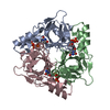
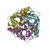
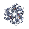
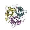
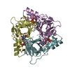
The biological assembly is a trimer and the asymmetric unit contains two such trimers. The first trimer is built from chain A, B and C and the second trimer is built from chains D, E and F
Mass: 18.015 Da / Num. of mol.: 427 / Source method: isolated from a natural source / Formula: H2O
-
Experimental details
-
Experiment
Experiment
Method: X-RAY DIFFRACTION / Number of used crystals: 1
-
Sample preparation
Crystal
Density Matthews: 2.18 Å3/Da / Density % sol: 44 %
Crystal grow
Temperature: 298 K / Method: vapor diffusion, hanging drop / pH: 7.5 Details: PEG 400, magnesium chloride, pH 7.5, VAPOR DIFFUSION, HANGING DROP, temperature 298K
-
Data collection
Diffraction
Mean temperature: 100 K
Diffraction source
Source: SYNCHROTRON / Site: MAX II / Beamline: I711 / Wavelength: 1.087 Å
Detector
Type: MARRESEARCH / Detector: CCD / Date: May 14, 2003
Radiation
Monochromator: Single asymmetrically cut Si(111) crystal with horizontal diffraction plane. The crystal is bendable for horizontal focusing Protocol: SINGLE WAVELENGTH / Monochromatic (M) / Laue (L): M / Scattering type: x-ray
Resolution: 1.8→1.84 Å / Rsym value: 0.238 / % possible all: 0.826
-
Processing
Software
Name
Classification
SHELX
modelbuilding
SHELXL-97
refinement
MAR345
datacollection
SCALEPACK
datascaling
Refinement
Method to determine structure: FOURIER SYNTHESIS / Resolution: 1.8→25 Å / Num. parameters: 38488 / Num. restraintsaints: 66686 / Cross valid method: FREE R / σ(F): 0 / Stereochemistry target values: Engh & Huber Details: this is a twinned structure. The twinning operator is (h,k,l) -> (h,k,-h-l) and the twinning fraction is 0.47. The R-factor is 16.44% and the R-free is 19.51% when this twinning operator is ...Details: this is a twinned structure. The twinning operator is (h,k,l) -> (h,k,-h-l) and the twinning fraction is 0.47. The R-factor is 16.44% and the R-free is 19.51% when this twinning operator is used. For F>4SIG(F) the corresponding values are 16.08% and 19.12%, respectively.
Rfactor
Num. reflection
% reflection
Selection details
Rfree
0.1951
4789
5.3 %
RANDOM
Rwork
0.1644
-
-
-
obs
0.1644
85935
90.1 %
-
all
-
90724
-
-
Refine analyze
Num. disordered residues: 10 / Occupancy sum hydrogen: 0 / Occupancy sum non hydrogen: 9577
Refinement step
Cycle: LAST / Resolution: 1.8→25 Å
Protein
Nucleic acid
Ligand
Solvent
Total
Num. atoms
9020
0
174
427
9621
Refine LS restraints
Refine-ID
Type
Dev ideal
X-RAY DIFFRACTION
s_bond_d
0.007
X-RAY DIFFRACTION
s_angle_d
0.021
X-RAY DIFFRACTION
s_similar_dist
0.045
X-RAY DIFFRACTION
s_from_restr_planes
0.028
X-RAY DIFFRACTION
s_zero_chiral_vol
0.033
X-RAY DIFFRACTION
s_non_zero_chiral_vol
0.04
X-RAY DIFFRACTION
s_anti_bump_dis_restr
0.006
X-RAY DIFFRACTION
s_rigid_bond_adp_cmpnt
0
X-RAY DIFFRACTION
s_similar_adp_cmpnt
0.058
X-RAY DIFFRACTION
s_approx_iso_adps
0
+
About Yorodumi
-
News
-
Feb 9, 2022. New format data for meta-information of EMDB entries
New format data for meta-information of EMDB entries
Version 3 of the EMDB header file is now the official format.
The previous official version 1.9 will be removed from the archive.
In the structure databanks used in Yorodumi, some data are registered as the other names, "COVID-19 virus" and "2019-nCoV". Here are the details of the virus and the list of structure data.
Jan 31, 2019. EMDB accession codes are about to change! (news from PDBe EMDB page)
EMDB accession codes are about to change! (news from PDBe EMDB page)
The allocation of 4 digits for EMDB accession codes will soon come to an end. Whilst these codes will remain in use, new EMDB accession codes will include an additional digit and will expand incrementally as the available range of codes is exhausted. The current 4-digit format prefixed with “EMD-” (i.e. EMD-XXXX) will advance to a 5-digit format (i.e. EMD-XXXXX), and so on. It is currently estimated that the 4-digit codes will be depleted around Spring 2019, at which point the 5-digit format will come into force.
The EM Navigator/Yorodumi systems omit the EMD- prefix.
Related info.:Q: What is EMD? / ID/Accession-code notation in Yorodumi/EM Navigator
Yorodumi is a browser for structure data from EMDB, PDB, SASBDB, etc.
This page is also the successor to EM Navigator detail page, and also detail information page/front-end page for Omokage search.
The word "yorodu" (or yorozu) is an old Japanese word meaning "ten thousand". "mi" (miru) is to see.
Related info.:EMDB / PDB / SASBDB / Comparison of 3 databanks / Yorodumi Search / Aug 31, 2016. New EM Navigator & Yorodumi / Yorodumi Papers / Jmol/JSmol / Function and homology information / Changes in new EM Navigator and Yorodumi
 Movie
Movie Controller


 Open data
 Basic information
 Components
 Keywords
 Function and homology information
 X-RAY DIFFRACTION /
  Authors
 Citation
 Structure visualization
 Downloads & links
 Other downloads
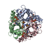


 PDBj
PDBj
 Assembly

 Mass: 24.305 Da / Num. of mol.: 6 / Source method: obtained synthetically / Formula: Mg
 Mass: 468.142 Da / Num. of mol.: 6 / Source method: obtained synthetically / Formula: C9H15N2O14P3
 Mass: 18.015 Da / Num. of mol.: 427 / Source method: isolated from a natural source / Formula: H2O
 Sample preparation
 / Beamline: I711 / Wavelength: 1.087 Å
 Processing