 Movie
Movie Controller

[English] 日本語
 Yorodumi
- PDB-2j4q: Crystal structure of a E138A Escherichia coli dCTP deaminase muta... -
+ Open data

- Basic information
| Entry | Database: PDB / ID: 2j4q | ||||||
|---|---|---|---|---|---|---|---|
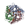
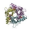
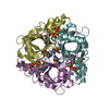
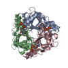
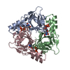
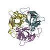
| Title | Crystal structure of a E138A Escherichia coli dCTP deaminase mutant enzyme in complex with dTTP | ||||||
 Components Components | DEOXYCYTIDINE TRIPHOSPHATE DEAMINASE | ||||||
 Keywords Keywords | HYDROLASE / NUCLEOTIDE METABOLISM / TRIMER / DCTP DEAMINASE | ||||||
| Function / homology |  Function and homology information Function and homology informationdCTP deaminase / dUTP biosynthetic process / dCTP deaminase activity / dUMP biosynthetic process / nucleobase-containing small molecule interconversion / dTTP biosynthetic process / protein homotrimerization / response to radiation / nucleotide binding / protein-containing complex ...dCTP deaminase / dUTP biosynthetic process / dCTP deaminase activity / dUMP biosynthetic process / nucleobase-containing small molecule interconversion / dTTP biosynthetic process / protein homotrimerization / response to radiation / nucleotide binding / protein-containing complex / identical protein binding / cytosol Similarity search - Function | ||||||
| Biological species |  | ||||||
| Method |  X-RAY DIFFRACTION / SYNCHROTRON / MOLECULAR REPLACEMENT / Resolution: 2.6 Å X-RAY DIFFRACTION / SYNCHROTRON / MOLECULAR REPLACEMENT / Resolution: 2.6 Å | ||||||
 Authors Authors | Johansson, E. / Thymark, M. / Bynck, J.H. / Fanoe, M. / Larsen, S. / Willemoes, M. | ||||||
 Citation Citation | Journal: FEBS J. / Year: 2007 Title: Regulation of Dctp Deaminase from Escherichia Coli by Nonallosteric Dttp Binding to an Inactive Form of the Enzyme Authors: Johansson, E. / Thymark, M. / Bynck, J.H. / Fanoe, M. / Larsen, S. / Willemoes, M. | ||||||
| History |
|
- Structure visualization
| Structure viewer | Molecule: MolmilJmol/JSmol |
|---|
- Downloads & links
-Download
| PDBx/mmCIF format | 2j4q.cif.gz | 83.1 KB | Display | PDBx/mmCIF format |
|---|---|---|---|---|
| PDB format | pdb2j4q.ent.gz | 61.5 KB | Display | PDB format |
| PDBx/mmJSON format | 2j4q.json.gz | Tree view | PDBx/mmJSON format | |
| Others |  Other downloads Other downloads |
-Validation report
| Arichive directory | https://data.pdbj.org/pub/pdb/validation_reports/j4/2j4qftp://data.pdbj.org/pub/pdb/validation_reports/j4/2j4q | HTTPS FTP |
|---|
-Related structure data
| Related structure data |  2j4hC  1xs1S S: Starting model for refinement C: citing same article ( |
|---|---|
| Similar structure data |


-Links
 PDBj
PDBj

- Assembly
| Deposited unit | 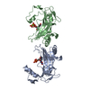
| |||||||||||||||||||||||||||||||||||||||||||||||||||||||||||||||||||||||||||||||||||||||||||||||||||||||||||||||||||||||||||||||||||||||||||||||||||
|---|---|---|---|---|---|---|---|---|---|---|---|---|---|---|---|---|---|---|---|---|---|---|---|---|---|---|---|---|---|---|---|---|---|---|---|---|---|---|---|---|---|---|---|---|---|---|---|---|---|---|---|---|---|---|---|---|---|---|---|---|---|---|---|---|---|---|---|---|---|---|---|---|---|---|---|---|---|---|---|---|---|---|---|---|---|---|---|---|---|---|---|---|---|---|---|---|---|---|---|---|---|---|---|---|---|---|---|---|---|---|---|---|---|---|---|---|---|---|---|---|---|---|---|---|---|---|---|---|---|---|---|---|---|---|---|---|---|---|---|---|---|---|---|---|---|---|---|---|
| 1 | 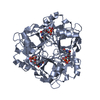
| |||||||||||||||||||||||||||||||||||||||||||||||||||||||||||||||||||||||||||||||||||||||||||||||||||||||||||||||||||||||||||||||||||||||||||||||||||
| 2 | 
| |||||||||||||||||||||||||||||||||||||||||||||||||||||||||||||||||||||||||||||||||||||||||||||||||||||||||||||||||||||||||||||||||||||||||||||||||||
| Unit cell |
| |||||||||||||||||||||||||||||||||||||||||||||||||||||||||||||||||||||||||||||||||||||||||||||||||||||||||||||||||||||||||||||||||||||||||||||||||||
| Components on special symmetry positions |
| |||||||||||||||||||||||||||||||||||||||||||||||||||||||||||||||||||||||||||||||||||||||||||||||||||||||||||||||||||||||||||||||||||||||||||||||||||
| Noncrystallographic symmetry (NCS) | NCS domain:
NCS domain segments: Ens-ID: 1 / Refine code: 1
|
