 Movie
Movie Controller

[English] 日本語
 Yorodumi
- PDB-1hqo: CRYSTAL STRUCTURE OF THE NITROGEN REGULATION FRAGMENT OF THE YEAS... -
+ Open data

- Basic information
| Entry | Database: PDB / ID: 1hqo | ||||||
|---|---|---|---|---|---|---|---|
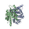
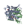
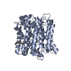
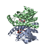
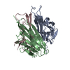
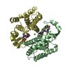
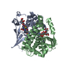
| Title | CRYSTAL STRUCTURE OF THE NITROGEN REGULATION FRAGMENT OF THE YEAST PRION PROTEIN URE2P | ||||||
 Components Components | URE2 PROTEIN | ||||||
 Keywords Keywords | SIGNALING PROTEIN / glutathione S-transferase superfamily fold | ||||||
| Function / homology |  Function and homology information Function and homology informationOxidoreductases; Acting on a sulfur group of donors; With a disulfide as acceptor / protein urmylation / glutathione peroxidase / regulation of nitrogen utilization / glutathione peroxidase activity / glutathione transferase activity / nitrate assimilation / phosphoprotein binding / transcription corepressor activity / intracellular protein localization ...Oxidoreductases; Acting on a sulfur group of donors; With a disulfide as acceptor / protein urmylation / glutathione peroxidase / regulation of nitrogen utilization / glutathione peroxidase activity / glutathione transferase activity / nitrate assimilation / phosphoprotein binding / transcription corepressor activity / intracellular protein localization / negative regulation of transcription by RNA polymerase II / cytoplasm Similarity search - Function | ||||||
| Biological species |  | ||||||
| Method |  X-RAY DIFFRACTION / SYNCHROTRON / MAD / Resolution: 2.3 Å X-RAY DIFFRACTION / SYNCHROTRON / MAD / Resolution: 2.3 Å | ||||||
 Authors Authors | Umland, T.C. / Taylor, K.L. / Rhee, S. / Wickner, R.B. / Davies, D.R. | ||||||
 Citation Citation | Journal: Proc.Natl.Acad.Sci.USA / Year: 2001 Title: The crystal structure of the nitrogen regulation fragment of the yeast prion protein Ure2p. Authors: Umland, T.C. / Taylor, K.L. / Rhee, S. / Wickner, R.B. / Davies, D.R. | ||||||
| History |
|
- Structure visualization
| Structure viewer | Molecule: MolmilJmol/JSmol |
|---|
- Downloads & links
-Download
| PDBx/mmCIF format | 1hqo.cif.gz | 107.5 KB | Display | PDBx/mmCIF format |
|---|---|---|---|---|
| PDB format | pdb1hqo.ent.gz | 83.2 KB | Display | PDB format |
| PDBx/mmJSON format | 1hqo.json.gz | Tree view | PDBx/mmJSON format | |
| Others |  Other downloads Other downloads |
-Validation report
| Arichive directory | https://data.pdbj.org/pub/pdb/validation_reports/hq/1hqoftp://data.pdbj.org/pub/pdb/validation_reports/hq/1hqo | HTTPS FTP |
|---|
-Related structure data
| Similar structure data |
|---|


-Links
 PDBj
PDBj


- Assembly
| Deposited unit | 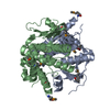
| ||||||||
|---|---|---|---|---|---|---|---|---|---|
| 1 |
| ||||||||
| Unit cell |
| ||||||||
| Details | The biological assembly is a dimer. The dimer was contained within the asymmetric unit. |
-Components
| #1: Protein | Mass: 29937.945 Da / Num. of mol.: 2 / Fragment: NITROGEN REGULATION FRAGMENT Source method: isolated from a genetically manipulated source Source: (gene. exp.) Gene: URE2 / Plasmid: PFLAG / Production host:  #2: Water | ChemComp-HOH / |  Mass: 18.015 Da / Num. of mol.: 204 / Source method: isolated from a natural source / Formula: H2O Mass: 18.015 Da / Num. of mol.: 204 / Source method: isolated from a natural source / Formula: H2OHas protein modification | Y | |
|---|
-Experimental details
-Experiment
| Experiment | Method: X-RAY DIFFRACTION / Number of used crystals: 1 |
|---|
- Sample preparation
| Crystal | Density Matthews: 2.79 Å3/Da / Density % sol: 55.84 % Description: The model was refined against the remote data set (0.9686 A). | ||||||||||||||||||||||||||||||||||||||||||
|---|---|---|---|---|---|---|---|---|---|---|---|---|---|---|---|---|---|---|---|---|---|---|---|---|---|---|---|---|---|---|---|---|---|---|---|---|---|---|---|---|---|---|---|
| Crystal grow | Temperature: 293 K / Method: vapor diffusion / pH: 8.8 Details: NaCl, Bicine, Tris-HCl, alpha-chymotrypsin, pH 8.8, VAPOR DIFFUSION, temperature 293K | ||||||||||||||||||||||||||||||||||||||||||
| Crystal grow | *PLUS pH: 8 | ||||||||||||||||||||||||||||||||||||||||||
| Components of the solutions | *PLUS
|
-Data collection
| Diffraction | Mean temperature: 100 K | ||||||||||||
|---|---|---|---|---|---|---|---|---|---|---|---|---|---|
| Diffraction source | Source: SYNCHROTRON / Site: NSLS  / Beamline: X9B / Wavelength: 0.9791, 0.9789, 0.9686 / Beamline: X9B / Wavelength: 0.9791, 0.9789, 0.9686 | ||||||||||||
| Detector | Type: ADSC QUANTUM 4 / Detector: CCD / Date: Nov 2, 2000 | ||||||||||||
| Radiation | Monochromator: Si monochomator / Protocol: MAD / Monochromatic (M) / Laue (L): M / Scattering type: x-ray | ||||||||||||
| Radiation wavelength |
| ||||||||||||
| Reflection | Resolution: 2.3→20 Å / Num. all: 29499 / Num. obs: 168715 / % possible obs: 97.6 % / Observed criterion σ(I): -3 / Redundancy: 5.7 % / Biso Wilson estimate: 50.9 Å2 / Rmerge(I) obs: 0.048 / Net I/σ(I): 15.4 | ||||||||||||
| Reflection shell | Resolution: 2.3→2.38 Å / Rmerge(I) obs: 0.32 / Num. unique all: 2941 / % possible all: 99.5 | ||||||||||||
| Reflection | *PLUS Lowest resolution: 20 Å / Num. obs: 29499 / Num. measured all: 168715 | ||||||||||||
| Reflection shell | *PLUS % possible obs: 99.5 % / Rmerge(I) obs: 0.32 |
- Processing
| Software |
| ||||||||||||||||||||||||||||||||||||
|---|---|---|---|---|---|---|---|---|---|---|---|---|---|---|---|---|---|---|---|---|---|---|---|---|---|---|---|---|---|---|---|---|---|---|---|---|---|
| Refinement | Method to determine structure: MAD / Resolution: 2.3→20 Å / Isotropic thermal model: Isotropic / Cross valid method: THROUGHOUT / σ(F): 2 / Stereochemistry target values: Engh & Huber
| ||||||||||||||||||||||||||||||||||||
| Displacement parameters | Biso mean: 55.6 Å2 | ||||||||||||||||||||||||||||||||||||
| Refine analyze |
| ||||||||||||||||||||||||||||||||||||
| Refinement step | Cycle: LAST / Resolution: 2.3→20 Å
| ||||||||||||||||||||||||||||||||||||
| Refine LS restraints |
| ||||||||||||||||||||||||||||||||||||
| LS refinement shell | Resolution: 2.3→2.38 Å / Total num. of bins used: 10
| ||||||||||||||||||||||||||||||||||||
| Software | *PLUS Name: CNS / Version: 1 / Classification: refinement | ||||||||||||||||||||||||||||||||||||
| Refinement | *PLUS Highest resolution: 2.3 Å / Lowest resolution: 20 Å / σ(F): 2 / % reflection Rfree: 5 % / Rfactor obs: 0.221 | ||||||||||||||||||||||||||||||||||||
| Solvent computation | *PLUS | ||||||||||||||||||||||||||||||||||||
| Displacement parameters | *PLUS Biso mean: 55.6 Å2 | ||||||||||||||||||||||||||||||||||||
| Refine LS restraints | *PLUS
| ||||||||||||||||||||||||||||||||||||
| LS refinement shell | *PLUS Highest resolution: 2.3 Å / Rfactor Rfree: 0.358 / Rfactor Rwork: 0.315 |
