 Movie
Movie Controller

[English] 日本語
 Yorodumi
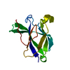
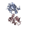
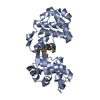
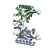
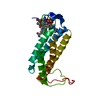
- PDB-6b15: Crystal structure of CBMbc (family CBM26) from Eubacterium rectal... -
+ Open data

- Basic information
| Entry | Database: PDB / ID: 6b15 | ||||||
|---|---|---|---|---|---|---|---|
| Title | Crystal structure of CBMbc (family CBM26) from Eubacterium rectale Amy13K | ||||||
 Components Components | Amy13K | ||||||
 Keywords Keywords | SUGAR BINDING PROTEIN / Carbohydrate Binding Module / Amylase / Starch / Gut Microbiome / Eubacterium rectale | ||||||
| Function / homology |  Function and homology information Function and homology informationhydrolase activity, acting on glycosyl bonds / carbohydrate metabolic process / membrane Similarity search - Function | ||||||
| Biological species |  Eubacterium rectale DSM 17629 (bacteria) Eubacterium rectale DSM 17629 (bacteria) | ||||||
| Method |  X-RAY DIFFRACTION / SYNCHROTRON / SAD / Resolution: 2.1 Å X-RAY DIFFRACTION / SYNCHROTRON / SAD / Resolution: 2.1 Å | ||||||
 Authors Authors | Cockburn, D.W. / Wawrzak, Z. / Perez Medina, K. / Koropatkin, N.M. | ||||||
 Citation Citation | Journal: Mol. Microbiol. / Year: 2018 Title: Novel carbohydrate binding modules in the surface anchored alpha-amylase of Eubacterium rectale provide a molecular rationale for the range of starches used by this organism in the human gut. Authors: Cockburn, D.W. / Suh, C. / Medina, K.P. / Duvall, R.M. / Wawrzak, Z. / Henrissat, B. / Koropatkin, N.M. | ||||||
| History |
|
- Structure visualization
| Structure viewer | Molecule: MolmilJmol/JSmol |
|---|
- Downloads & links
-Download
| PDBx/mmCIF format | 6b15.cif.gz | 311.3 KB | Display | PDBx/mmCIF format |
|---|---|---|---|---|
| PDB format | pdb6b15.ent.gz | 255 KB | Display | PDB format |
| PDBx/mmJSON format | 6b15.json.gz | Tree view | PDBx/mmJSON format | |
| Others |  Other downloads Other downloads |
-Validation report
| Arichive directory | https://data.pdbj.org/pub/pdb/validation_reports/b1/6b15ftp://data.pdbj.org/pub/pdb/validation_reports/b1/6b15 | HTTPS FTP |
|---|
-Related structure data


-Links
 PDBj
PDBj

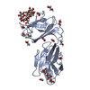
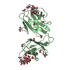
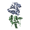
- Assembly
| Deposited unit | 
| |||||||||
|---|---|---|---|---|---|---|---|---|---|---|
| 1 | 
| |||||||||
| 2 | 
| |||||||||
| 3 | 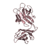
| |||||||||
| 4 | 
| |||||||||
| Unit cell |
| |||||||||
| Components on special symmetry positions |
|
-Components
| #1: Protein | Mass: 22953.947 Da / Num. of mol.: 4 Source method: isolated from a genetically manipulated source Source: (gene. exp.) Eubacterium rectale DSM 17629 (bacteria)Production host: #2: Chemical |   Mass: 62.068 Da / Num. of mol.: 3 / Source method: obtained synthetically / Formula: C2H6O2 Mass: 62.068 Da / Num. of mol.: 3 / Source method: obtained synthetically / Formula: C2H6O2#3: Water | ChemComp-HOH / |  Mass: 18.015 Da / Num. of mol.: 730 / Source method: isolated from a natural source / Formula: H2O Mass: 18.015 Da / Num. of mol.: 730 / Source method: isolated from a natural source / Formula: H2O |
|---|
-Experimental details
-Experiment
| Experiment | Method: X-RAY DIFFRACTION / Number of used crystals: 1 |
|---|
- Sample preparation
| Crystal | Density Matthews: 4.14 Å3/Da / Density % sol: 70.31 % |
|---|---|
| Crystal grow | Temperature: 295 K / Method: vapor diffusion, hanging drop / pH: 7 / Details: 60% Tacsimate, 0.1 M Bis-Tris Propane, pH 7.0 |
-Data collection
| Diffraction | Mean temperature: 80 K |
|---|---|
| Diffraction source | Source: SYNCHROTRON / Site: APS  / Beamline: 21-ID-D / Wavelength: 0.979 Å / Beamline: 21-ID-D / Wavelength: 0.979 Å |
| Detector | Type: MARMOSAIC 225 mm CCD / Detector: CCD / Date: Apr 18, 2015 |
| Radiation | Protocol: SINGLE WAVELENGTH / Monochromatic (M) / Laue (L): M / Scattering type: x-ray |
| Radiation wavelength | Wavelength: 0.979 Å / Relative weight: 1 |
| Reflection | Resolution: 2.1→36.02 Å / Num. obs: 86483 / % possible obs: 98.46 % / Redundancy: 6.5 % / CC1/2: 0.995 / Rmerge(I) obs: 0.09175 / Rpim(I) all: 0.03852 / Net I/σ(I): 7.86 |
| Reflection shell | Resolution: 2.1→2.175 Å / Redundancy: 6.4 % / Rmerge(I) obs: 0.5615 / Mean I/σ(I) obs: 2.03 / Num. unique obs: 8664 / CC1/2: 0.832 / Rpim(I) all: 0.237 / % possible all: 99.38 |
- Processing
| Software |
| |||||||||||||||||||||||||||||||||||||||||||||||||||||||||||||||||||||||||||||||||||||||||||||||||||||||||
|---|---|---|---|---|---|---|---|---|---|---|---|---|---|---|---|---|---|---|---|---|---|---|---|---|---|---|---|---|---|---|---|---|---|---|---|---|---|---|---|---|---|---|---|---|---|---|---|---|---|---|---|---|---|---|---|---|---|---|---|---|---|---|---|---|---|---|---|---|---|---|---|---|---|---|---|---|---|---|---|---|---|---|---|---|---|---|---|---|---|---|---|---|---|---|---|---|---|---|---|---|---|---|---|---|---|---|
| Refinement | Method to determine structure: SAD / Resolution: 2.1→36.02 Å / SU ML: 0.27 / Cross valid method: FREE R-VALUE / σ(F): 1.36 / Phase error: 24.8
| |||||||||||||||||||||||||||||||||||||||||||||||||||||||||||||||||||||||||||||||||||||||||||||||||||||||||
| Solvent computation | Shrinkage radii: 0.9 Å / VDW probe radii: 1.11 Å | |||||||||||||||||||||||||||||||||||||||||||||||||||||||||||||||||||||||||||||||||||||||||||||||||||||||||
| Refinement step | Cycle: LAST / Resolution: 2.1→36.02 Å
| |||||||||||||||||||||||||||||||||||||||||||||||||||||||||||||||||||||||||||||||||||||||||||||||||||||||||
| Refine LS restraints |
| |||||||||||||||||||||||||||||||||||||||||||||||||||||||||||||||||||||||||||||||||||||||||||||||||||||||||
| LS refinement shell |
|
