Resolution: 2.54→46.42 Å / Num. obs: 31756 / % possible obs: 93.4 % / Redundancy: 6.49 % / Biso Wilson estimate: 54.685 Å2 / Rmerge(I) obs: 0.091 / Net I/σ(I): 15.62
Reflection shell
Diffraction-ID: 1
Resolution (Å)
Redundancy (%)
Rmerge(I) obs
Mean I/σ(I) obs
Num. measured obs
Num. unique obs
% possible all
2.54-2.63
2
0.39
2
5582
2793
86.1
2.63-2.74
2
0.307
2.6
6275
3111
93.3
2.74-2.86
2.9
0.367
3.6
8681
2983
94.8
2.86-3.01
8.1
0.474
5.5
26092
3203
100
3.01-3.2
8.2
0.322
7.5
26906
3271
100
3.2-3.44
8.2
0.181
12.4
26028
3176
100
3.44-3.79
8.1
0.106
18.6
26807
3290
100
3.79-4.33
8.2
0.065
28
26449
3242
100
4.33-5.44
8.1
0.058
30.8
26459
3284
100
5.44-46.4
7.9
0.038
40.4
26908
3403
99.8
-
Phasing
Phasing
Method: MAD
-
Processing
Software
Name
Version
Classification
NB
MolProbity
3beta29
modelbuilding
REFMAC
5.2.0019
refinement
XSCALE
datascaling
PDB_EXTRACT
1.701
dataextraction
XDS
datareduction
SHELX
phasing
autoSHARP
phasing
Refinement
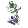
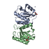
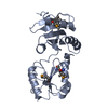
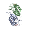
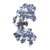
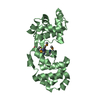
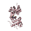
Method to determine structure: MAD / Resolution: 2.55→46.4 Å / Cor.coef. Fo:Fc: 0.948 / Cor.coef. Fo:Fc free: 0.914 / SU B: 19.51 / SU ML: 0.191 / TLS residual ADP flag: LIKELY RESIDUAL / Cross valid method: THROUGHOUT / σ(F): 0 / ESU R: 0.343 / ESU R Free: 0.257 Stereochemistry target values: MAXIMUM LIKELIHOOD WITH PHASES Details: 1. HYDROGENS HAVE BEEN ADDED IN THE RIDING POSITIONS. 2. THERE ARE THREE MONOMERS IN THE ASSYMMETRIC UNIT. CHAIN A AND B ARE VERY SIMILAR TO EACH OTHER. CHAIN C DIFFERS FROM A/B DUE TO INTER- ...Details: 1. HYDROGENS HAVE BEEN ADDED IN THE RIDING POSITIONS. 2. THERE ARE THREE MONOMERS IN THE ASSYMMETRIC UNIT. CHAIN A AND B ARE VERY SIMILAR TO EACH OTHER. CHAIN C DIFFERS FROM A/B DUE TO INTER-DOMAIN MOVEMENT. THE MOVEMENT IN THE MONOMER C CAN BE DESCRIBED BY ROTATION OF ~23 DEGREE AROUND RESIDUES 33-34,110-126. 3. THERE ARE SIGNIFICANT DISORDER IN THE DOMAIN C38-148. THE FOLLOWING RESIDUES ARE NOT MODELED DESPITE PRESENCE DISORDERED DENSITY: C81, C115-121, C130-133. 4. A MET-INHIBITION PROTOCOL WAS USED FOR SELENOMETHIONINE INCORPORATION DURING PROTEIN EXPRESSION. THE OCCUPANCY OF THE SE ATOMS IN THE MSE RESIDUES WAS REDUCED TO 0.7 TO ACCOUNT FOR THE REDUCED SCATTERING POWER DUE TO PARTIAL S-MET INCORPORATION. 5. ATOM RECORD CONTAINS RESIDUAL B FACTORS ONLY.
Rfactor
Num. reflection
% reflection
Selection details
Rfree
0.246
1606
5.1 %
RANDOM
Rwork
0.196
-
-
-
obs
0.198
31729
98 %
-
Solvent computation
Ion probe radii: 0.8 Å / Shrinkage radii: 0.8 Å / VDW probe radii: 1.2 Å / Solvent model: BABINET MODEL WITH MASK
Displacement parameters
Biso mean: 57.814 Å2
Baniso -1
Baniso -2
Baniso -3
1-
1.61 Å2
0.81 Å2
0 Å2
2-
-
1.61 Å2
0 Å2
3-
-
-
-2.42 Å2
Refinement step
Cycle: LAST / Resolution: 2.55→46.4 Å
Protein
Nucleic acid
Ligand
Solvent
Total
Num. atoms
4815
0
0
40
4855
Refine LS restraints
Refine-ID
Type
Dev ideal
Dev ideal target
Number
X-RAY DIFFRACTION
r_bond_refined_d
0.014
0.022
4911
X-RAY DIFFRACTION
r_bond_other_d
0.001
0.02
3259
X-RAY DIFFRACTION
r_angle_refined_deg
1.418
1.953
6648
X-RAY DIFFRACTION
r_angle_other_deg
0.93
3
7924
X-RAY DIFFRACTION
r_dihedral_angle_1_deg
5.808
5
631
X-RAY DIFFRACTION
r_dihedral_angle_2_deg
35.165
23.385
195
X-RAY DIFFRACTION
r_dihedral_angle_3_deg
16.788
15
787
X-RAY DIFFRACTION
r_dihedral_angle_4_deg
16.676
15
27
X-RAY DIFFRACTION
r_chiral_restr
0.073
0.2
748
X-RAY DIFFRACTION
r_gen_planes_refined
0.005
0.02
5512
X-RAY DIFFRACTION
r_gen_planes_other
0.001
0.02
1027
X-RAY DIFFRACTION
r_nbd_refined
0.236
0.2
1148
X-RAY DIFFRACTION
r_nbd_other
0.181
0.2
3160
X-RAY DIFFRACTION
r_nbtor_refined
0.194
0.2
2436
X-RAY DIFFRACTION
r_nbtor_other
0.088
0.2
2509
X-RAY DIFFRACTION
r_xyhbond_nbd_refined
0.173
0.2
66
X-RAY DIFFRACTION
r_xyhbond_nbd_other
0.026
0.2
1
X-RAY DIFFRACTION
r_symmetry_vdw_refined
0.152
0.2
18
X-RAY DIFFRACTION
r_symmetry_vdw_other
0.238
0.2
59
X-RAY DIFFRACTION
r_symmetry_hbond_refined
0.197
0.2
11
X-RAY DIFFRACTION
r_mcbond_it
1.693
3
3223
X-RAY DIFFRACTION
r_mcbond_other
0.367
3
1310
X-RAY DIFFRACTION
r_mcangle_it
2.885
5
4977
X-RAY DIFFRACTION
r_scbond_it
5.484
8
1963
X-RAY DIFFRACTION
r_scangle_it
7.528
11
1671
Refine LS restraints NCS
Refine-ID: X-RAY DIFFRACTION
Ens-ID
Dom-ID
Auth asym-ID
Number
Type
Rms dev position (Å)
Weight position
1
1
A
1453
LOOSEPOSITIONAL
0.64
5
1
2
B
1453
LOOSEPOSITIONAL
0.61
5
1
3
C
1453
LOOSEPOSITIONAL
0.55
5
1
1
A
1453
LOOSETHERMAL
3.46
10
1
2
B
1453
LOOSETHERMAL
2.68
10
1
3
C
1453
LOOSETHERMAL
3.54
10
2
1
A
1213
LOOSEPOSITIONAL
0.44
5
2
1
A
1213
LOOSETHERMAL
2.78
10
LS refinement shell
Resolution: 2.545→2.611 Å / Total num. of bins used: 20
Rfactor
Num. reflection
% reflection
Rfree
0.395
119
-
Rwork
0.306
2045
-
obs
-
2164
91.23 %
Refinement TLS params.
Method: refined / Refine-ID: X-RAY DIFFRACTION
ID
L11 (°2)
L12 (°2)
L13 (°2)
L22 (°2)
L23 (°2)
L33 (°2)
S11 (Å °)
S12 (Å °)
S13 (Å °)
S21 (Å °)
S22 (Å °)
S23 (Å °)
S31 (Å °)
S32 (Å °)
S33 (Å °)
T11 (Å2)
T12 (Å2)
T13 (Å2)
T22 (Å2)
T23 (Å2)
T33 (Å2)
Origin x (Å)
Origin y (Å)
Origin z (Å)
1
4.1316
-2.0025
0.8348
6.1036
0.2097
3.7913
0.0781
-0.4822
-0.2207
0.2274
-0.1573
0.453
0.1806
-0.6436
0.0792
-0.4389
0.0264
-0.075
-0.1065
-0.0266
-0.2739
21.8206
34.6505
17.8407
2
4.914
0.6737
0.5237
2.0402
0.2856
3.6895
-0.1426
0.0744
0.2318
-0.0854
0.0889
-0.0227
0.2049
0.1875
0.0538
-0.331
0.1233
-0.0917
-0.3599
-0.0748
-0.2712
9.4419
41.1646
-6.144
3
2.4046
-0.122
-1.0639
10.468
5.5701
6.1484
0.1351
-0.0615
-0.0039
0.7735
0.394
-1.2611
0.677
0.2956
-0.5291
-0.2694
-0.0351
-0.1202
-0.2879
-0.1508
-0.1026
-6.948
55.092
17.438
4
2.1228
0.9008
2.2614
3.3581
2.8389
5.8668
0.1387
0.1494
-0.0709
0.1211
-0.1743
0.0327
0.4214
-0.1505
0.0356
-0.2119
-0.0307
-0.0004
-0.3315
0.0036
-0.3138
-7.5165
77.6512
35.3978
5
3.5976
0.0287
0.1165
4.4077
1.4915
5.2197
0.1865
0.1567
-0.0045
-0.5441
-0.0431
0.3351
-0.292
-0.2062
-0.1434
-0.2071
0.0878
0.0101
-0.2932
0.0286
-0.2448
9.9717
61.5029
61.0275
6
3.9203
-1.4376
-3.2842
3.4352
0.8023
11.0406
0.1761
0.0528
0.4338
0.1139
0.2567
-0.7855
-0.791
1.2574
-0.4329
0.1979
0.0283
-0.0479
0.171
-0.177
-0.032
23.4167
57.9875
38.8825
Refinement TLS group
Refine-ID: X-RAY DIFFRACTION / Selection: ALL
ID
Refine TLS-ID
Auth asym-ID
Label asym-ID
Auth seq-ID
Label seq-ID
1
1
A
A
0 - 37
12 - 49
2
1
A
A
149 - 217
161 - 229
3
2
A
A
38 - 148
50 - 160
4
3
B
B
1 - 37
13 - 49
5
3
B
B
149 - 217
161 - 229
6
4
B
B
38 - 148
50 - 160
7
5
C
C
1 - 37
13 - 49
8
5
C
C
149 - 220
161 - 232
9
6
C
C
38 - 148
50 - 160
+
About Yorodumi
-
News
-
Feb 9, 2022. New format data for meta-information of EMDB entries
New format data for meta-information of EMDB entries
Version 3 of the EMDB header file is now the official format.
The previous official version 1.9 will be removed from the archive.
In the structure databanks used in Yorodumi, some data are registered as the other names, "COVID-19 virus" and "2019-nCoV". Here are the details of the virus and the list of structure data.
Jan 31, 2019. EMDB accession codes are about to change! (news from PDBe EMDB page)
EMDB accession codes are about to change! (news from PDBe EMDB page)
The allocation of 4 digits for EMDB accession codes will soon come to an end. Whilst these codes will remain in use, new EMDB accession codes will include an additional digit and will expand incrementally as the available range of codes is exhausted. The current 4-digit format prefixed with “EMD-” (i.e. EMD-XXXX) will advance to a 5-digit format (i.e. EMD-XXXXX), and so on. It is currently estimated that the 4-digit codes will be depleted around Spring 2019, at which point the 5-digit format will come into force.
The EM Navigator/Yorodumi systems omit the EMD- prefix.
Related info.:Q: What is EMD? / ID/Accession-code notation in Yorodumi/EM Navigator
Yorodumi is a browser for structure data from EMDB, PDB, SASBDB, etc.
This page is also the successor to EM Navigator detail page, and also detail information page/front-end page for Omokage search.
The word "yorodu" (or yorozu) is an old Japanese word meaning "ten thousand". "mi" (miru) is to see.
Related info.:EMDB / PDB / SASBDB / Comparison of 3 databanks / Yorodumi Search / Aug 31, 2016. New EM Navigator & Yorodumi / Yorodumi Papers / Jmol/JSmol / Function and homology information / Changes in new EM Navigator and Yorodumi
 Movie
Movie Controller

 Yorodumi
 Open data
 Basic information
 Components
 Keywords
 Function and homology information
 Bacillus halodurans (bacteria)
 X-RAY DIFFRACTION /
  Authors
 Citation
 Structure visualization
 Downloads & links
 Other downloads


 PDBj
PDBj Assembly
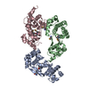
 Mass: 18.015 Da / Num. of mol.: 40 / Source method: isolated from a natural source / Formula: H2O
 Sample preparation
 / Beamline: 8.2.2 / Wavelength: 1.0163, 0.9798
 Processing