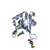
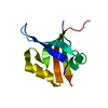
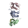
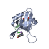
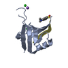
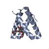
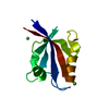
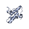
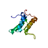
Entry Database : PDB / ID : 4nxpTitle Crystal Structure of Free T-cell Lymphoma Invasion and Metastasis-1 PDZ Domain Quadruple Mutant (QM) T-lymphoma invasion and metastasis-inducing protein 1 Keywords / / / / / / Function / homology Function Domain/homology Component
/ / / / / / / / / / / / / / / / / / / / / / / / / / / / / / / / / / / / / / / / / / / / / / / / / / / / / / / / / / / / / / / / / / / / / / / / / / / / / / / Biological species Homo sapiens (human)Method / / Resolution : 2.3 Å Authors Liu, X. / Speckhard, D.C. / Shepherd, T.R. / Hengel, S.R. / Fuentes, E.J. Journal : Structure / Year : 2016Title : Distinct Roles for Conformational Dynamics in Protein-Ligand Interactions.Authors : Liu, X. / Speckhard, D.C. / Shepherd, T.R. / Sun, Y.J. / Hengel, S.R. / Yu, L. / Fowler, C.A. / Gakhar, L. / Fuentes, E.J. History Deposition Dec 9, 2013 Deposition site / Processing site Revision 1.0 May 13, 2015 Provider / Type Revision 1.1 Feb 15, 2017 Group Revision 1.2 Dec 21, 2022 Group / Category / struct_ref_seq_difItem / _database_2.pdbx_database_accession / _struct_ref_seq_dif.detailsRevision 1.3 Sep 20, 2023 Group / Refinement descriptionCategory / chem_comp_bond / pdbx_initial_refinement_model
Show all Show less
 Movie
Movie Controller

 Yorodumi
 Open data
 Basic information
 Components
 Keywords
 Function and homology information
 Homo sapiens (human)
 X-RAY DIFFRACTION /
  Authors
 Citation
 Structure visualization
 Downloads & links
 Other downloads
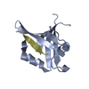


 PDBj
PDBj


 Assembly
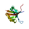

 Mass: 18.015 Da / Num. of mol.: 47 / Source method: isolated from a natural source / Formula: H2O
 Sample preparation
 Processing