- PDB-2eaq: Crystal structure of PDZ domain of KIAA0858 (LIM), MS0793 from Ho... -
+
Open data
ID or keywords:
Loading...
-
Basic information
Entry
Database: PDB / ID: 2eaq
Title
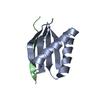
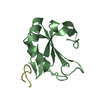
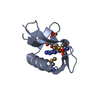
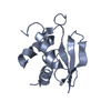
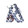
Crystal structure of PDZ domain of KIAA0858 (LIM), MS0793 from Homo sapiens
Components
LIM domain only protein 7
Keywords
METAL BINDING PROTEIN / conserved hypothetical protein / Structural Genomics / NPPSFA / National Project on Protein Structural and Functional Analyses / RIKEN Structural Genomics/Proteomics Initiative / RSGI
Function / homology
Function and homology information
regulation of signaling / regulation of cell adhesion / ubiquitin ligase complex / ubiquitin-protein transferase activity / Signaling by ALK fusions and activated point mutants / nuclear envelope / Antigen processing: Ubiquitination & Proteasome degradation / Neddylation / apical plasma membrane / protein ubiquitination ...regulation of signaling / regulation of cell adhesion / ubiquitin ligase complex / ubiquitin-protein transferase activity / Signaling by ALK fusions and activated point mutants / nuclear envelope / Antigen processing: Ubiquitination & Proteasome degradation / Neddylation / apical plasma membrane / protein ubiquitination / focal adhesion / cell surface / positive regulation of transcription by RNA polymerase II / mitochondrion / metal ion binding / nucleus / cytoplasm / cytosol Similarity search - Function
LIM domain only protein 7 / Domain of unknown function DUF4757 / Domain of unknown function (DUF4757) / Smooth muscle protein/calponin / LIM zinc-binding domain signature. / LIM domain / Zinc-binding domain present in Lin-11, Isl-1, Mec-3. / Zinc finger, LIM-type / LIM domain profile. / Calponin homology domain ...LIM domain only protein 7 / Domain of unknown function DUF4757 / Domain of unknown function (DUF4757) / Smooth muscle protein/calponin / LIM zinc-binding domain signature. / LIM domain / Zinc-binding domain present in Lin-11, Isl-1, Mec-3. / Zinc finger, LIM-type / LIM domain profile. / Calponin homology domain / Calponin homology (CH) domain / Calponin homology domain / CH domain superfamily / Calponin homology (CH) domain profile. / PDZ domain / Pdz3 Domain / PDZ domain / PDZ domain profile. / Domain present in PSD-95, Dlg, and ZO-1/2. / PDZ domain / PDZ superfamily / Roll / Mainly Beta Similarity search - Domain/homology
In the structure databanks used in Yorodumi, some data are registered as the other names, "COVID-19 virus" and "2019-nCoV". Here are the details of the virus and the list of structure data.
Jan 31, 2019. EMDB accession codes are about to change! (news from PDBe EMDB page)
EMDB accession codes are about to change! (news from PDBe EMDB page)
The allocation of 4 digits for EMDB accession codes will soon come to an end. Whilst these codes will remain in use, new EMDB accession codes will include an additional digit and will expand incrementally as the available range of codes is exhausted. The current 4-digit format prefixed with “EMD-” (i.e. EMD-XXXX) will advance to a 5-digit format (i.e. EMD-XXXXX), and so on. It is currently estimated that the 4-digit codes will be depleted around Spring 2019, at which point the 5-digit format will come into force.
The EM Navigator/Yorodumi systems omit the EMD- prefix.
Related info.:Q: What is EMD? / ID/Accession-code notation in Yorodumi/EM Navigator
Yorodumi is a browser for structure data from EMDB, PDB, SASBDB, etc.
This page is also the successor to EM Navigator detail page, and also detail information page/front-end page for Omokage search.
The word "yorodu" (or yorozu) is an old Japanese word meaning "ten thousand". "mi" (miru) is to see.
Related info.:EMDB / PDB / SASBDB / Comparison of 3 databanks / Yorodumi Search / Aug 31, 2016. New EM Navigator & Yorodumi / Yorodumi Papers / Jmol/JSmol / Function and homology information / Changes in new EM Navigator and Yorodumi
 Movie
Movie Controller

 Yorodumi
 Open data
 Basic information
 Components
 Keywords
 Function and homology information
 Homo sapiens (human)
 X-RAY DIFFRACTION /
  Authors
 Citation
 Structure visualization
 Downloads & links
 Other downloads
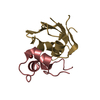


 PDBj
PDBj Assembly
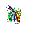

 Mass: 58.693 Da / Num. of mol.: 2 / Source method: obtained synthetically / Formula: Ni
 Mass: 18.015 Da / Num. of mol.: 137 / Source method: isolated from a natural source / Formula: H2O
 Sample preparation
 / Beamline: BL-5A / Wavelength: 0.9794, 0.9796, 0.9644
 Processing