 Movie
Movie Controller

[English] 日本語
 Yorodumi
- PDB-4hlx: The crystal structure of the DNA binding domain of vIRF-1 from th... -
+ Open data

- Basic information
| Entry | Database: PDB / ID: 4hlx | ||||||
|---|---|---|---|---|---|---|---|
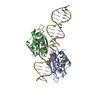
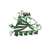
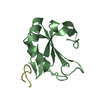
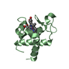
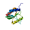
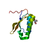
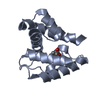
| Title | The crystal structure of the DNA binding domain of vIRF-1 from the oncogenic KSHV | ||||||
 Components Components | K9 | ||||||
 Keywords Keywords | DNA BINDING PROTEIN / helix-turn-helix | ||||||
| Function / homology |  Function and homology information Function and homology informationimmune system process / DNA-binding transcription factor activity, RNA polymerase II-specific / RNA polymerase II cis-regulatory region sequence-specific DNA binding Similarity search - Function | ||||||
| Biological species |   Human herpesvirus 8 Human herpesvirus 8 | ||||||
| Method |  X-RAY DIFFRACTION / SYNCHROTRON / MOLECULAR REPLACEMENT / Resolution: 2.379 Å X-RAY DIFFRACTION / SYNCHROTRON / MOLECULAR REPLACEMENT / Resolution: 2.379 Å | ||||||
 Authors Authors | Hew, K. / Venkatachalam, R. | ||||||
 Citation Citation | Journal: Nucleic Acids Res. / Year: 2013 Title: The crystal structure of the DNA-binding domain of vIRF-1 from the oncogenic KSHV reveals a conserved fold for DNA binding and reinforces its role as a transcription factor. Authors: Hew, K. / Dahlroth, S.L. / Venkatachalam, R. / Nasertorabi, F. / Lim, B.T. / Cornvik, T. / Nordlund, P. | ||||||
| History |
|
- Structure visualization
| Structure viewer | Molecule: MolmilJmol/JSmol |
|---|
- Downloads & links
-Download
| PDBx/mmCIF format | 4hlx.cif.gz | 93.9 KB | Display | PDBx/mmCIF format |
|---|---|---|---|---|
| PDB format | pdb4hlx.ent.gz | 72 KB | Display | PDB format |
| PDBx/mmJSON format | 4hlx.json.gz | Tree view | PDBx/mmJSON format | |
| Others |  Other downloads Other downloads |
-Validation report
| Arichive directory | https://data.pdbj.org/pub/pdb/validation_reports/hl/4hlxftp://data.pdbj.org/pub/pdb/validation_reports/hl/4hlx | HTTPS FTP |
|---|
-Related structure data


-Links
 PDBj
PDBj

- Assembly
| Deposited unit | 
| ||||||||
|---|---|---|---|---|---|---|---|---|---|
| 1 | 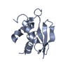
| ||||||||
| 2 | 
| ||||||||
| 3 | 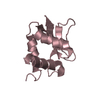
| ||||||||
| 4 | 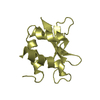
| ||||||||
| Unit cell |
|
-Components
| #1: Protein | Mass: 15309.481 Da / Num. of mol.: 4 / Fragment: DNA binding domain, UNP RESIDUES 88-196 Source method: isolated from a genetically manipulated source Source: (gene. exp.) Human herpesvirus 8 / Gene: ORF K9, vIRF, vIRF-1 / Plasmid: pNIC28-Bsa4 / Production host:  #2: Water | ChemComp-HOH / |  Mass: 18.015 Da / Num. of mol.: 117 / Source method: isolated from a natural source / Formula: H2O Mass: 18.015 Da / Num. of mol.: 117 / Source method: isolated from a natural source / Formula: H2OHas protein modification | Y | |
|---|
-Experimental details
-Experiment
| Experiment | Method: X-RAY DIFFRACTION / Number of used crystals: 1 |
|---|
- Sample preparation
| Crystal | Density Matthews: 1.98 Å3/Da / Density % sol: 37.75 % |
|---|---|
| Crystal grow | Temperature: 277 K / Method: vapor diffusion, sitting drop / pH: 7 Details: 0.1 M HEPES pH 7, 27% Jeffamine-ED 2001, 10 mM ATP, VAPOR DIFFUSION, SITTING DROP, temperature 277K |
-Data collection
| Diffraction | Mean temperature: 110 K |
|---|---|
| Diffraction source | Source: SYNCHROTRON / Site: NSRRC  / Beamline: BL13B1 / Wavelength: 0.96722 Å / Beamline: BL13B1 / Wavelength: 0.96722 Å |
| Detector | Type: ADSC QUANTUM 315r / Detector: CCD / Date: Nov 28, 2011 |
| Radiation | Monochromator: Rh Coated Mirror / Protocol: SINGLE WAVELENGTH / Monochromatic (M) / Laue (L): M / Scattering type: x-ray |
| Radiation wavelength | Wavelength: 0.96722 Å / Relative weight: 1 |
| Reflection | Resolution: 2.379→23.292 Å / Num. all: 18285 / Num. obs: 18285 / % possible obs: 92.5 % / Redundancy: 6.6 % |
| Reflection shell | Highest resolution: 2.38 Å / Redundancy: 6.6 % / Num. unique all: 18285 / % possible all: 92.58 |
- Processing
| Software |
| ||||||||||||||||||||||||||||||||||||||||||
|---|---|---|---|---|---|---|---|---|---|---|---|---|---|---|---|---|---|---|---|---|---|---|---|---|---|---|---|---|---|---|---|---|---|---|---|---|---|---|---|---|---|---|---|
| Refinement | Method to determine structure: MOLECULAR REPLACEMENT / Resolution: 2.379→23.292 Å / SU ML: 0.36 / σ(F): 1.42 / Phase error: 32.12 / Stereochemistry target values: ML
| ||||||||||||||||||||||||||||||||||||||||||
| Solvent computation | Shrinkage radii: 0.8 Å / VDW probe radii: 1.1 Å / Solvent model: FLAT BULK SOLVENT MODEL | ||||||||||||||||||||||||||||||||||||||||||
| Refinement step | Cycle: LAST / Resolution: 2.379→23.292 Å
| ||||||||||||||||||||||||||||||||||||||||||
| Refine LS restraints |
| ||||||||||||||||||||||||||||||||||||||||||
| LS refinement shell | Refine-ID: X-RAY DIFFRACTION / Total num. of bins used: 6
|
