 Movie
Movie Controller


+ Open data

- Basic information
| Entry | Database: PDB / ID: 7al1 | ||||||
|---|---|---|---|---|---|---|---|
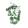
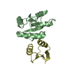
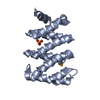
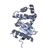
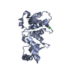
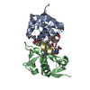
| Title | Cell division protein SepF from Methanobrevibacter smithii | ||||||
 Components Components | Cell division protein SepF | ||||||
 Keywords Keywords | CELL CYCLE / FtsZ-binding protein Membrane-binding protein | ||||||
| Function / homology | SepF-like superfamily / Cell division protein SepF Function and homology information Function and homology information | ||||||
| Biological species |  Methanobrevibacter smithii (archaea) Methanobrevibacter smithii (archaea) | ||||||
| Method |  X-RAY DIFFRACTION / SYNCHROTRON / MOLECULAR REPLACEMENT / Resolution: 1.4 Å X-RAY DIFFRACTION / SYNCHROTRON / MOLECULAR REPLACEMENT / Resolution: 1.4 Å | ||||||
 Authors Authors | Sogues, A. / wehenkel, A.M. / Alzari, P.M. | ||||||
| Funding support |  France, 1items France, 1items
| ||||||
 Citation Citation | Journal: Nat Commun / Year: 2021 Title: SepF is the FtsZ anchor in archaea, with features of an ancestral cell division system. Authors: Pende, N. / Sogues, A. / Megrian, D. / Sartori-Rupp, A. / England, P. / Palabikyan, H. / Rittmann, S.K.R. / Grana, M. / Wehenkel, A.M. / Alzari, P.M. / Gribaldo, S. | ||||||
| History |
|
- Structure visualization
| Structure viewer | Molecule: MolmilJmol/JSmol |
|---|
- Downloads & links
-Download
| PDBx/mmCIF format | 7al1.cif.gz | 78.8 KB | Display | PDBx/mmCIF format |
|---|---|---|---|---|
| PDB format | pdb7al1.ent.gz | 49.5 KB | Display | PDB format |
| PDBx/mmJSON format | 7al1.json.gz | Tree view | PDBx/mmJSON format | |
| Others |  Other downloads Other downloads |
-Validation report
| Arichive directory | https://data.pdbj.org/pub/pdb/validation_reports/al/7al1ftp://data.pdbj.org/pub/pdb/validation_reports/al/7al1 | HTTPS FTP |
|---|
-Related structure data
| Related structure data |  7al2C 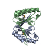 3zieS S: Starting model for refinement C: citing same article ( |
|---|---|
| Similar structure data |


-Links
 PDBj
PDBj- Assembly
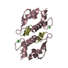
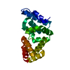
| Deposited unit | 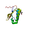
| ||||||||||||
|---|---|---|---|---|---|---|---|---|---|---|---|---|---|
| 1 | 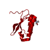
| ||||||||||||
| Unit cell |
| ||||||||||||
| Components on special symmetry positions |
|
-Components
| #1: Protein | Mass: 10851.482 Da / Num. of mol.: 1 Source method: isolated from a genetically manipulated source Source: (gene. exp.) Methanobrevibacter smithii (strain ATCC 35061 / DSM 861 / OCM 144 / PS) (archaea)Strain: ATCC 35061 / DSM 861 / OCM 144 / PS / Gene: Msm_0406 / Production host:  |
|---|---|
| #2: Chemical | ChemComp-TRS /   Mass: 122.143 Da / Num. of mol.: 1 / Source method: obtained synthetically / Formula: C4H12NO3 / Comment: pH buffer*YM Mass: 122.143 Da / Num. of mol.: 1 / Source method: obtained synthetically / Formula: C4H12NO3 / Comment: pH buffer*YM |
| #3: Water | ChemComp-HOH /  Mass: 18.015 Da / Num. of mol.: 57 / Source method: isolated from a natural source / Formula: H2O Mass: 18.015 Da / Num. of mol.: 57 / Source method: isolated from a natural source / Formula: H2O |
| Has ligand of interest | N |
-Experimental details
-Experiment
| Experiment | Method: X-RAY DIFFRACTION / Number of used crystals: 1 |
|---|
- Sample preparation
| Crystal | Density Matthews: 2.02 Å3/Da / Density % sol: 39.06 % |
|---|---|
| Crystal grow | Temperature: 291 K / Method: vapor diffusion, sitting drop / pH: 8.5 / Details: 0.1 M TRIS, pH 8.5, 30% PEG 10K |
-Data collection
| Diffraction | Mean temperature: 100 K / Serial crystal experiment: N |
|---|---|
| Diffraction source | Source: SYNCHROTRON / Site: SOLEIL / Beamline: PROXIMA 1 / Wavelength: 0.9875 Å |
| Detector | Type: DECTRIS EIGER X 16M / Detector: PIXEL / Date: Nov 8, 2019 |
| Radiation | Protocol: SINGLE WAVELENGTH / Monochromatic (M) / Laue (L): M / Scattering type: x-ray |
| Radiation wavelength | Wavelength: 0.9875 Å / Relative weight: 1 |
| Reflection | Resolution: 1.4→45.95 Å / Num. obs: 17704 / % possible obs: 99.9 % / Redundancy: 19.6 % / Biso Wilson estimate: 20.19 Å2 / Rmerge(I) obs: 0.056 / Net I/σ(I): 27.1 |
| Reflection shell | Resolution: 1.4→1.42 Å / Rmerge(I) obs: 1.029 / Mean I/σ(I) obs: 3.4 / Num. unique obs: 846 |
- Processing
| Software |
| |||||||||||||||||||||||||||||||||||||||||||||||||
|---|---|---|---|---|---|---|---|---|---|---|---|---|---|---|---|---|---|---|---|---|---|---|---|---|---|---|---|---|---|---|---|---|---|---|---|---|---|---|---|---|---|---|---|---|---|---|---|---|---|---|
| Refinement | Method to determine structure: MOLECULAR REPLACEMENT Starting model: pdb code 3ZIE Resolution: 1.4→34.97 Å / SU ML: 0.1155 / Cross valid method: FREE R-VALUE / σ(F): 1.34 / Phase error: 25.0207 Stereochemistry target values: GeoStd + Monomer Library + CDL v1.2
| |||||||||||||||||||||||||||||||||||||||||||||||||
| Solvent computation | Shrinkage radii: 0.9 Å / VDW probe radii: 1.11 Å / Solvent model: FLAT BULK SOLVENT MODEL | |||||||||||||||||||||||||||||||||||||||||||||||||
| Displacement parameters | Biso mean: 26.38 Å2 | |||||||||||||||||||||||||||||||||||||||||||||||||
| Refinement step | Cycle: LAST / Resolution: 1.4→34.97 Å
| |||||||||||||||||||||||||||||||||||||||||||||||||
| Refine LS restraints |
| |||||||||||||||||||||||||||||||||||||||||||||||||
| LS refinement shell |
| |||||||||||||||||||||||||||||||||||||||||||||||||
| Refinement TLS params. | Method: refined / Origin x: -4.29269921737 Å / Origin y: -22.3702062537 Å / Origin z: 8.34574490984 Å
|
