 Movie
Movie Controller

[English] 日本語
 Yorodumi
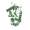
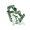
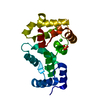
- PDB-2het: Non-myristoylated bovine recoverin (truncated at C-terminus) with... -
+ Open data

- Basic information
| Entry | Database: PDB / ID: 2het | ||||||
|---|---|---|---|---|---|---|---|
| Title | Non-myristoylated bovine recoverin (truncated at C-terminus) with calcium bound to EF-hand 3 | ||||||
 Components Components | Recoverin | ||||||
 Keywords Keywords | METAL BINDING PROTEIN / Recoverin / EF-hand / helix-loop-helix / calcium binding | ||||||
| Function / homology |  Function and homology information Function and homology informationInactivation, recovery and regulation of the phototransduction cascade / regulation of calcium ion transport / phototransduction / regulation of signal transduction / photoreceptor outer segment / photoreceptor inner segment / visual perception / perikaryon / calcium ion binding / cytosol Similarity search - Function | ||||||
| Biological species |  | ||||||
| Method |  X-RAY DIFFRACTION / SYNCHROTRON / MOLECULAR REPLACEMENT / Resolution: 3 Å X-RAY DIFFRACTION / SYNCHROTRON / MOLECULAR REPLACEMENT / Resolution: 3 Å | ||||||
 Authors Authors | Weiergraber, O.H. / Granzin, J. | ||||||
 Citation Citation | Journal: J.Biol.Chem. / Year: 2006 Title: Tuning of a neuronal calcium sensor. Authors: Weiergraber, O.H. / Senin, I.I. / Zernii, E.Y. / Churumova, V.A. / Kovaleva, N.A. / Nazipova, A.A. / Permyakov, S.E. / Permyakov, E.A. / Philippov, P.P. / Granzin, J. / Koch, K.W. | ||||||
| History |
|
- Structure visualization
| Structure viewer | Molecule: MolmilJmol/JSmol |
|---|
- Downloads & links
-Download
| PDBx/mmCIF format | 2het.cif.gz | 141.5 KB | Display | PDBx/mmCIF format |
|---|---|---|---|---|
| PDB format | pdb2het.ent.gz | 111.8 KB | Display | PDB format |
| PDBx/mmJSON format | 2het.json.gz | Tree view | PDBx/mmJSON format | |
| Others |  Other downloads Other downloads |
-Validation report
| Arichive directory | https://data.pdbj.org/pub/pdb/validation_reports/he/2hetftp://data.pdbj.org/pub/pdb/validation_reports/he/2het | HTTPS FTP |
|---|
-Related structure data
| Related structure data | 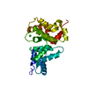 1omrS S: Starting model for refinement |
|---|---|
| Similar structure data |


-Links
 PDBj
PDBj


- Assembly
| Deposited unit | 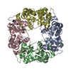
| ||||||||
|---|---|---|---|---|---|---|---|---|---|
| 1 | 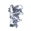
| ||||||||
| 2 | 
| ||||||||
| 3 | 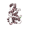
| ||||||||
| 4 | 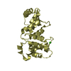
| ||||||||
| 5 |
| ||||||||
| Unit cell |
|
-Components
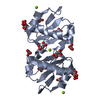
| #1: Protein | Mass: 21748.322 Da / Num. of mol.: 4 Source method: isolated from a genetically manipulated source Source: (gene. exp.)  #2: Chemical | ChemComp-CA /   Mass: 40.078 Da / Num. of mol.: 4 / Source method: obtained synthetically / Formula: Ca Mass: 40.078 Da / Num. of mol.: 4 / Source method: obtained synthetically / Formula: Ca |
|---|
-Experimental details
-Experiment
| Experiment | Method: X-RAY DIFFRACTION / Number of used crystals: 1 |
|---|
- Sample preparation
| Crystal | Density Matthews: 2.45 Å3/Da / Density % sol: 49.71 % |
|---|---|
| Crystal grow | Temperature: 290 K / Method: vapor diffusion, hanging drop / pH: 7 Details: 2.4 M sodium malonate, 2 mM calcium chloride, pH 7.0, VAPOR DIFFUSION, HANGING DROP, temperature 290K |
-Data collection
| Diffraction | Mean temperature: 100 K |
|---|---|
| Diffraction source | Source: SYNCHROTRON / Site: ESRF  / Beamline: ID14-1 / Wavelength: 0.934 Å / Beamline: ID14-1 / Wavelength: 0.934 Å |
| Detector | Type: ADSC QUANTUM 4 / Detector: CCD / Date: Feb 25, 2006 / Details: Monochromators, mirrors |
| Radiation | Monochromator: Diamond, germanium / Protocol: SINGLE WAVELENGTH / Monochromatic (M) / Laue (L): M / Scattering type: x-ray |
| Radiation wavelength | Wavelength: 0.934 Å / Relative weight: 1 |
| Reflection | Resolution: 3→70 Å / Num. all: 14380 / Num. obs: 14380 / % possible obs: 84.8 % / Observed criterion σ(F): 2 / Observed criterion σ(I): 2 / Redundancy: 2.4 % / Biso Wilson estimate: 89.4 Å2 / Rmerge(I) obs: 0.044 / Rsym value: 0.044 / Net I/σ(I): 11.3 |
| Reflection shell | Resolution: 3→3.16 Å / Redundancy: 2.2 % / Rmerge(I) obs: 0.234 / Mean I/σ(I) obs: 1.7 / Num. unique all: 2072 / Rsym value: 0.234 / % possible all: 84.1 |
- Processing
| Software |
| ||||||||||||||||||||
|---|---|---|---|---|---|---|---|---|---|---|---|---|---|---|---|---|---|---|---|---|---|
| Refinement | Method to determine structure: MOLECULAR REPLACEMENT Starting model: PDB ENTRY 1OMR Resolution: 3→70 Å / Isotropic thermal model: Isotropic / Cross valid method: THROUGHOUT / σ(F): 0 / Stereochemistry target values: Engh & Huber
| ||||||||||||||||||||
| Displacement parameters | Biso mean: 77.4 Å2
| ||||||||||||||||||||
| Refine analyze |
| ||||||||||||||||||||
| Refinement step | Cycle: LAST / Resolution: 3→70 Å
| ||||||||||||||||||||
| Refine LS restraints |
|
