- PDB-4k1g: Structure of E. coli Nfo(Endo IV)-H69A mutant bound to a cleaved ... -
+
Open data
ID or keywords:
Loading...
-
Basic information
Entry
Database: PDB / ID: 4k1g
Title
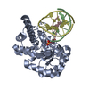
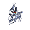
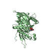
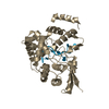
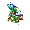
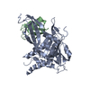
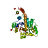
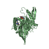
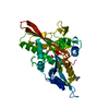
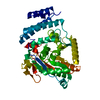
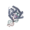
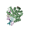
Structure of E. coli Nfo(Endo IV)-H69A mutant bound to a cleaved DNA duplex containing a alphadA:T basepair
Components
DNA (5'-D(*CP*GP*TP*CP*GP*TP*CP*GP*TP*GP*GP*AP*CP*GP*C)-3')
DNA (5'-D(*GP*CP*GP*TP*CP*C)-3')
DNA (5'-D(P*AP*CP*GP*AP*CP*GP*AP*CP*G)-3')
Endonuclease 4
Keywords
HYDROLASE/DNA / DNA endonuclease IV / HYDROLASE-DNA complex
Function / homology
Function and homology information
deoxyribonuclease IV / deoxyribonuclease IV (phage-T4-induced) activity / phosphoric diester hydrolase activity / 3'-5'-DNA exonuclease activity / phosphatase activity / DNA-(apurinic or apyrimidinic site) endonuclease activity / base-excision repair / DNA repair / DNA binding / zinc ion binding / cytosol Similarity search - Function
AP endonucleases family 2 signature 1. / AP endonucleases family 2 signature 2. / AP endonucleases family 2 signature 3. / AP endonuclease 2, zinc binding site / AP endonucleases family 2 profile. / AP endonuclease family 2 / AP endonuclease 2 / Divalent-metal-dependent TIM barrel enzymes / Xylose isomerase-like, TIM barrel domain / Xylose isomerase-like TIM barrel ...AP endonucleases family 2 signature 1. / AP endonucleases family 2 signature 2. / AP endonucleases family 2 signature 3. / AP endonuclease 2, zinc binding site / AP endonucleases family 2 profile. / AP endonuclease family 2 / AP endonuclease 2 / Divalent-metal-dependent TIM barrel enzymes / Xylose isomerase-like, TIM barrel domain / Xylose isomerase-like TIM barrel / Xylose isomerase-like superfamily / TIM Barrel / Alpha-Beta Barrel / Alpha Beta Similarity search - Domain/homology
A: Endonuclease 4 B: Endonuclease 4 E: DNA (5'-D(*GP*CP*GP*TP*CP*C)-3') F: DNA (5'-D(*CP*GP*TP*CP*GP*TP*CP*GP*TP*GP*GP*AP*CP*GP*C)-3') H: DNA (5'-D(P*AP*CP*GP*AP*CP*GP*AP*CP*G)-3') M: DNA (5'-D(*GP*CP*GP*TP*CP*C)-3') N: DNA (5'-D(*CP*GP*TP*CP*GP*TP*CP*GP*TP*GP*GP*AP*CP*GP*C)-3') O: DNA (5'-D(P*AP*CP*GP*AP*CP*GP*AP*CP*G)-3') hetero molecules
A: Endonuclease 4 E: DNA (5'-D(*GP*CP*GP*TP*CP*C)-3') F: DNA (5'-D(*CP*GP*TP*CP*GP*TP*CP*GP*TP*GP*GP*AP*CP*GP*C)-3') H: DNA (5'-D(P*AP*CP*GP*AP*CP*GP*AP*CP*G)-3') hetero molecules
B: Endonuclease 4 M: DNA (5'-D(*GP*CP*GP*TP*CP*C)-3') N: DNA (5'-D(*CP*GP*TP*CP*GP*TP*CP*GP*TP*GP*GP*AP*CP*GP*C)-3') O: DNA (5'-D(P*AP*CP*GP*AP*CP*GP*AP*CP*G)-3') hetero molecules
AUTHORS STATE THAT THE QUATERNARY STRUCTURE IS A PROTEIN MONOMER-DOUBLE STRANDED DNA COMPLEX. ONE OF THE DNA STRANDS IS CLEAVED BETWEEN THE BASE 306 AND THE BASE 307.
-
Components
-
Protein , 1 types, 2 molecules AB
#1: Protein
Endonuclease4 / Endodeoxyribonuclease IV / Endonuclease IV
Mass: 31451.430 Da / Num. of mol.: 2 / Mutation: H69A Source method: isolated from a genetically manipulated source Source: (gene. exp.) Escherichia coli (E. coli) / Strain: K12 / Gene: nfo, b2159, JW2146 / Production host: Escherichia coli (E. coli) / References: UniProt: P0A6C1, deoxyribonuclease IV
-
DNA chain , 3 types, 6 molecules EMFNHO
#2: DNA chain
DNA (5'-D(*GP*CP*GP*TP*CP*C)-3')
Mass: 1785.193 Da / Num. of mol.: 2 / Source method: obtained synthetically
#3: DNA chain
DNA (5'-D(*CP*GP*TP*CP*GP*TP*CP*GP*TP*GP*GP*AP*CP*GP*C)-3')
Mass: 4601.971 Da / Num. of mol.: 2 / Source method: obtained synthetically
#4: DNA chain
DNA (5'-D(P*AP*CP*GP*AP*CP*GP*AP*CP*G)-3')
Mass: 2749.826 Da / Num. of mol.: 2 / Source method: obtained synthetically
Mass: 18.015 Da / Num. of mol.: 610 / Source method: isolated from a natural source / Formula: H2O
-
Details
Sequence details
CHAINS E AND H, AND CHAINS M AND O ARE FORM THE SAME DNA STRAND WHICH IS CLEAVED BETWEEN THE BASE ...CHAINS E AND H, AND CHAINS M AND O ARE FORM THE SAME DNA STRAND WHICH IS CLEAVED BETWEEN THE BASE 306 AND THE BASE 307.
-
Experimental details
-
Experiment
Experiment
Method: X-RAY DIFFRACTION / Number of used crystals: 1
-
Sample preparation
Crystal
Density Matthews: 2.79 Å3/Da / Density % sol: 55.94 %
Crystal grow
Temperature: 293 K / Method: vapor diffusion, hanging drop / pH: 8 Details: PEG 4000, Tris HCl, pH 8.0, VAPOR DIFFUSION, HANGING DROP, temperature 293K
In the structure databanks used in Yorodumi, some data are registered as the other names, "COVID-19 virus" and "2019-nCoV". Here are the details of the virus and the list of structure data.
Jan 31, 2019. EMDB accession codes are about to change! (news from PDBe EMDB page)
EMDB accession codes are about to change! (news from PDBe EMDB page)
The allocation of 4 digits for EMDB accession codes will soon come to an end. Whilst these codes will remain in use, new EMDB accession codes will include an additional digit and will expand incrementally as the available range of codes is exhausted. The current 4-digit format prefixed with “EMD-” (i.e. EMD-XXXX) will advance to a 5-digit format (i.e. EMD-XXXXX), and so on. It is currently estimated that the 4-digit codes will be depleted around Spring 2019, at which point the 5-digit format will come into force.
The EM Navigator/Yorodumi systems omit the EMD- prefix.
Related info.:Q: What is EMD? / ID/Accession-code notation in Yorodumi/EM Navigator
Yorodumi is a browser for structure data from EMDB, PDB, SASBDB, etc.
This page is also the successor to EM Navigator detail page, and also detail information page/front-end page for Omokage search.
The word "yorodu" (or yorozu) is an old Japanese word meaning "ten thousand". "mi" (miru) is to see.
Related info.:EMDB / PDB / SASBDB / Comparison of 3 databanks / Yorodumi Search / Aug 31, 2016. New EM Navigator & Yorodumi / Yorodumi Papers / Jmol/JSmol / Function and homology information / Changes in new EM Navigator and Yorodumi
 Movie
Movie Controller

 Yorodumi
 Open data
 Basic information
 Components
 Keywords
 Function and homology information
 X-RAY DIFFRACTION /
  Authors
 Citation
 Structure visualization
 Downloads & links
 Other downloads
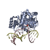

 PDBj
PDBj


 Assembly
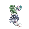


 Mass: 65.409 Da / Num. of mol.: 5 / Source method: obtained synthetically / Formula: Zn
 Mass: 106.120 Da / Num. of mol.: 2 / Source method: obtained synthetically / Formula: C4H10O3
 Sample preparation
 / Beamline: PROXIMA 1 / Wavelength: 0.9
  Processing