 Movie
Movie Controller

[English] 日本語
 Yorodumi
- PDB-5ohn: Crystal structure of USP30 in covalent complex with ubiquitin pro... -
+ Open data

- Basic information
| Entry | Database: PDB / ID: 5ohn | ||||||
|---|---|---|---|---|---|---|---|
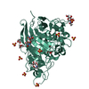
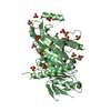
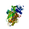
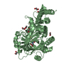
| Title | Crystal structure of USP30 in covalent complex with ubiquitin propargylamide (low resolution) | ||||||
 Components Components |
| ||||||
 Keywords Keywords | HYDROLASE / Deubiquitinase / DUB / Ubiquitin / USP | ||||||
| Function / homology |  Function and homology information Function and homology informationprotein K6-linked deubiquitination / protein K11-linked deubiquitination / deubiquitinase activity / autophagy of mitochondrion / pexophagy / hypothalamus gonadotrophin-releasing hormone neuron development / mitochondrial fusion / female meiosis I / positive regulation of protein monoubiquitination / peroxisomal membrane ...protein K6-linked deubiquitination / protein K11-linked deubiquitination / deubiquitinase activity / autophagy of mitochondrion / pexophagy / hypothalamus gonadotrophin-releasing hormone neuron development / mitochondrial fusion / female meiosis I / positive regulation of protein monoubiquitination / peroxisomal membrane / fat pad development / mitochondrion transport along microtubule / seminiferous tubule development / female gonad development / negative regulation of mitophagy / male meiosis I / positive regulation of intrinsic apoptotic signaling pathway by p53 class mediator / protein deubiquitination / energy homeostasis / neuron projection morphogenesis / regulation of proteasomal protein catabolic process / Maturation of protein E / Maturation of protein E / ER Quality Control Compartment (ERQC) / Myoclonic epilepsy of Lafora / FLT3 signaling by CBL mutants / IRAK2 mediated activation of TAK1 complex / Alpha-protein kinase 1 signaling pathway / Glycogen synthesis / IRAK1 recruits IKK complex / IRAK1 recruits IKK complex upon TLR7/8 or 9 stimulation / Prevention of phagosomal-lysosomal fusion / Endosomal Sorting Complex Required For Transport (ESCRT) / Membrane binding and targetting of GAG proteins / Regulation of TBK1, IKKε (IKBKE)-mediated activation of IRF3, IRF7 / Negative regulation of FLT3 / PTK6 Regulates RTKs and Their Effectors AKT1 and DOK1 / Regulation of TBK1, IKKε-mediated activation of IRF3, IRF7 upon TLR3 ligation / IRAK2 mediated activation of TAK1 complex upon TLR7/8 or 9 stimulation / Constitutive Signaling by NOTCH1 HD Domain Mutants / NOTCH2 Activation and Transmission of Signal to the Nucleus / TICAM1,TRAF6-dependent induction of TAK1 complex / TICAM1-dependent activation of IRF3/IRF7 / APC/C:Cdc20 mediated degradation of Cyclin B / regulation of neuron apoptotic process / Downregulation of ERBB4 signaling / APC-Cdc20 mediated degradation of Nek2A / Regulation of FZD by ubiquitination / p75NTR recruits signalling complexes / InlA-mediated entry of Listeria monocytogenes into host cells / TRAF6 mediated IRF7 activation in TLR7/8 or 9 signaling / NF-kB is activated and signals survival / TRAF6-mediated induction of TAK1 complex within TLR4 complex / Regulation of pyruvate metabolism / Pexophagy / Downregulation of ERBB2:ERBB3 signaling / Regulation of innate immune responses to cytosolic DNA / NRIF signals cell death from the nucleus / Regulation of PTEN localization / positive regulation of protein ubiquitination / VLDLR internalisation and degradation / Activated NOTCH1 Transmits Signal to the Nucleus / Synthesis of active ubiquitin: roles of E1 and E2 enzymes / Translesion synthesis by REV1 / TICAM1, RIP1-mediated IKK complex recruitment / Regulation of BACH1 activity / Translesion synthesis by POLK / JNK (c-Jun kinases) phosphorylation and activation mediated by activated human TAK1 / InlB-mediated entry of Listeria monocytogenes into host cell / MAP3K8 (TPL2)-dependent MAPK1/3 activation / Activation of IRF3, IRF7 mediated by TBK1, IKKε (IKBKE) / Downregulation of TGF-beta receptor signaling / Translesion synthesis by POLI / Josephin domain DUBs / Gap-filling DNA repair synthesis and ligation in GG-NER / IKK complex recruitment mediated by RIP1 / PINK1-PRKN Mediated Mitophagy / regulation of mitochondrial membrane potential / TGF-beta receptor signaling in EMT (epithelial to mesenchymal transition) / TNFR1-induced NF-kappa-B signaling pathway / Regulation of activated PAK-2p34 by proteasome mediated degradation / TCF dependent signaling in response to WNT / Regulation of NF-kappa B signaling / activated TAK1 mediates p38 MAPK activation / Autodegradation of Cdh1 by Cdh1:APC/C / APC/C:Cdc20 mediated degradation of Securin / NOTCH3 Activation and Transmission of Signal to the Nucleus / N-glycan trimming in the ER and Calnexin/Calreticulin cycle / Regulation of signaling by CBL / Negative regulators of DDX58/IFIH1 signaling / Asymmetric localization of PCP proteins / Fanconi Anemia Pathway / Ubiquitin-dependent degradation of Cyclin D / Negative regulation of FGFR3 signaling / Peroxisomal protein import / Deactivation of the beta-catenin transactivating complex / SCF-beta-TrCP mediated degradation of Emi1 / NIK-->noncanonical NF-kB signaling / AUF1 (hnRNP D0) binds and destabilizes mRNA / TNFR2 non-canonical NF-kB pathway Similarity search - Function | ||||||
| Biological species |  Homo sapiens (human) Homo sapiens (human) | ||||||
| Method |  X-RAY DIFFRACTION / SYNCHROTRON / MOLECULAR REPLACEMENT / Resolution: 3.6 Å X-RAY DIFFRACTION / SYNCHROTRON / MOLECULAR REPLACEMENT / Resolution: 3.6 Å | ||||||
 Authors Authors | Gersch, M. / Komander, D. | ||||||
| Funding support |  United Kingdom, 1items United Kingdom, 1items
| ||||||
 Citation Citation | Journal: Nat. Struct. Mol. Biol. / Year: 2017 Title: Mechanism and regulation of the Lys6-selective deubiquitinase USP30. Authors: Gersch, M. / Gladkova, C. / Schubert, A.F. / Michel, M.A. / Maslen, S. / Komander, D. | ||||||
| History |
|
- Structure visualization
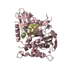
| Structure viewer | Molecule: MolmilJmol/JSmol |
|---|
- Downloads & links
-Download
| PDBx/mmCIF format | 5ohn.cif.gz | 161.9 KB | Display | PDBx/mmCIF format |
|---|---|---|---|---|
| PDB format | pdb5ohn.ent.gz | 124.3 KB | Display | PDB format |
| PDBx/mmJSON format | 5ohn.json.gz | Tree view | PDBx/mmJSON format | |
| Others |  Other downloads Other downloads |
-Validation report
| Arichive directory | https://data.pdbj.org/pub/pdb/validation_reports/oh/5ohnftp://data.pdbj.org/pub/pdb/validation_reports/oh/5ohn | HTTPS FTP |
|---|
-Related structure data
| Related structure data | 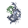 5ohkSC  5ohpC S: Starting model for refinement C: citing same article ( |
|---|---|
| Similar structure data |


-Links
 PDBj
PDBj


- Assembly
| Deposited unit | 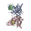
| ||||||||
|---|---|---|---|---|---|---|---|---|---|
| 1 | 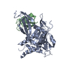
| ||||||||
| 2 | 
| ||||||||
| Unit cell |
|
-Components
| #1: Protein | Mass: 42582.031 Da / Num. of mol.: 2 / Fragment: UNP residues 64-360,UNP residues 432-502 Mutation: construct 8 (F348D, M350D, I353E, L358S, L359N, G360A),construct 8 (F348D, M350D, I353E, L358S, L359N, G360A) Source method: isolated from a genetically manipulated source Source: (gene. exp.) Homo sapiens (human) / Gene: USP30 / Plasmid: MG-26-21 / Production host:  #2: Protein | Mass: 8558.857 Da / Num. of mol.: 2 Source method: isolated from a genetically manipulated source Source: (gene. exp.) Homo sapiens (human) / Gene: UBB / Production host: #3: Chemical |   Mass: 65.409 Da / Num. of mol.: 2 / Source method: obtained synthetically / Formula: Zn Mass: 65.409 Da / Num. of mol.: 2 / Source method: obtained synthetically / Formula: ZnHas protein modification | Y | |
|---|
-Experimental details
-Experiment
| Experiment | Method: X-RAY DIFFRACTION / Number of used crystals: 1 |
|---|
- Sample preparation
| Crystal | Density Matthews: 4.43 Å3/Da / Density % sol: 72.26 % |
|---|---|
| Crystal grow | Temperature: 291 K / Method: vapor diffusion, sitting drop / pH: 8 Details: 20% (w/v) PAA 5100 Na, 100 mM Hepes pH 8.0, 2.5% (v/v) glycerol |
-Data collection
| Diffraction | Mean temperature: 100 K | |||||||||||||||
|---|---|---|---|---|---|---|---|---|---|---|---|---|---|---|---|---|
| Diffraction source | Source: SYNCHROTRON / Site: Diamond / Beamline: I04-1 / Wavelength: 0.92819 Å | |||||||||||||||
| Detector | Type: DECTRIS PILATUS 6M-F / Detector: PIXEL / Date: Jan 17, 2016 | |||||||||||||||
| Radiation | Protocol: SINGLE WAVELENGTH / Monochromatic (M) / Laue (L): M / Scattering type: x-ray | |||||||||||||||
| Radiation wavelength | Wavelength: 0.92819 Å / Relative weight: 1 | |||||||||||||||
| Reflection twin |
| |||||||||||||||
| Reflection | Resolution: 3.6→157.47 Å / Num. obs: 20776 / % possible obs: 99.3 % / Redundancy: 7 % / CC1/2: 0.997 / Rmerge(I) obs: 0.124 / Rpim(I) all: 0.072 / Net I/σ(I): 10 | |||||||||||||||
| Reflection shell | Resolution: 3.6→3.94 Å / Redundancy: 7.2 % / Rmerge(I) obs: 0.9 / Mean I/σ(I) obs: 1.9 / Num. unique obs: 4948 / CC1/2: 0.593 / Rpim(I) all: 0.523 / % possible all: 99.6 |
- Processing
| Software |
| ||||||||||||||||||||||||||||||||||||||||||||||||||||||||
|---|---|---|---|---|---|---|---|---|---|---|---|---|---|---|---|---|---|---|---|---|---|---|---|---|---|---|---|---|---|---|---|---|---|---|---|---|---|---|---|---|---|---|---|---|---|---|---|---|---|---|---|---|---|---|---|---|---|
| Refinement | Method to determine structure: MOLECULAR REPLACEMENT Starting model: 5OHK Resolution: 3.6→157.473 Å / Cross valid method: THROUGHOUT / σ(F): 201.05 / Phase error: 22.68 / Stereochemistry target values: TWIN_LSQ_F
| ||||||||||||||||||||||||||||||||||||||||||||||||||||||||
| Solvent computation | Shrinkage radii: 0.9 Å / VDW probe radii: 1.11 Å / Solvent model: FLAT BULK SOLVENT MODEL | ||||||||||||||||||||||||||||||||||||||||||||||||||||||||
| Refinement step | Cycle: LAST / Resolution: 3.6→157.473 Å
| ||||||||||||||||||||||||||||||||||||||||||||||||||||||||
| Refine LS restraints |
| ||||||||||||||||||||||||||||||||||||||||||||||||||||||||
| LS refinement shell |
|
