 Movie
Movie Controller

[English] 日本語
 Yorodumi
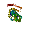
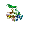
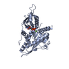
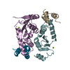
- PDB-1k30: Crystal Structure Analysis of Squash (Cucurbita moschata) glycero... -
+ Open data

- Basic information
| Entry | Database: PDB / ID: 1k30 | ||||||
|---|---|---|---|---|---|---|---|
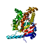
| Title | Crystal Structure Analysis of Squash (Cucurbita moschata) glycerol-3-phosphate (1)-acyltransferase | ||||||
 Components Components | glycerol-3-phosphate acyltransferase | ||||||
 Keywords Keywords | TRANSFERASE / Four-helix bundle | ||||||
| Function / homology |  Function and homology information Function and homology information: / glycerol-3-phosphate 1-O-acyltransferase / glycerol-3-phosphate O-acyltransferase activity / phosphatidylglycerol biosynthetic process / CDP-diacylglycerol biosynthetic process / chloroplast stroma Similarity search - Function | ||||||
| Biological species |  Cucurbita moschata (crookneck pumpkin) Cucurbita moschata (crookneck pumpkin) | ||||||
| Method |  X-RAY DIFFRACTION / MIR / Resolution: 1.9 Å X-RAY DIFFRACTION / MIR / Resolution: 1.9 Å | ||||||
 Authors Authors | Turnbull, A.P. / Rafferty, J.B. / Sedelnikova, S.E. / Slabas, A.R. / Schierer, T.P. / Kroon, J.T. / Simon, J.W. / Fawcett, T. / Nishida, I. / Murata, N. / Rice, D.W. | ||||||
 Citation Citation | Journal: Structure / Year: 2001 Title: Analysis of the structure, substrate specificity, and mechanism of squash glycerol-3-phosphate (1)-acyltransferase. Authors: Turnbull, A.P. / Rafferty, J.B. / Sedelnikova, S.E. / Slabas, A.R. / Schierer, T.P. / Kroon, J.T. / Simon, J.W. / Fawcett, T. / Nishida, I. / Murata, N. / Rice, D.W. #1: Journal: Acta Crystallogr.,Sect.D / Year: 2001Title: Crystallization and preliminary X-ray analysis of the glycerol-3-phosphate 1-acyltransferase from squash (Cucurbita moschata). Authors: Turnbull, A.P. / Rafferty, J.B. / Sedelnikova, S.E. / Slabas, A.R. / Schierer, T.P. / Kroon, J.T. / Nishida, I. / Murata, N. / Simon, J.W. / Rice, D.W. | ||||||
| History |
|
- Structure visualization
| Structure viewer | Molecule: MolmilJmol/JSmol |
|---|
- Downloads & links
-Download
| PDBx/mmCIF format | 1k30.cif.gz | 91.6 KB | Display | PDBx/mmCIF format |
|---|---|---|---|---|
| PDB format | pdb1k30.ent.gz | 69.7 KB | Display | PDB format |
| PDBx/mmJSON format | 1k30.json.gz | Tree view | PDBx/mmJSON format | |
| Others |  Other downloads Other downloads |
-Validation report
| Arichive directory | https://data.pdbj.org/pub/pdb/validation_reports/k3/1k30ftp://data.pdbj.org/pub/pdb/validation_reports/k3/1k30 | HTTPS FTP |
|---|
-Related structure data
| Similar structure data |
|---|


-Links
 PDBj
PDBj- Assembly
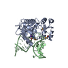
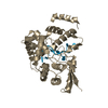
| Deposited unit | 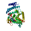
| ||||||||
|---|---|---|---|---|---|---|---|---|---|
| 1 |
| ||||||||
| Unit cell |
|
-Components
| #1: Protein | Mass: 40968.555 Da / Num. of mol.: 1 Source method: isolated from a genetically manipulated source Source: (gene. exp.) Cucurbita moschata (crookneck pumpkin) / Gene: PLSB / Plasmid: pET24a / Species (production host): Escherichia coli / Production host:  References: UniProt: P10349, glycerol-3-phosphate 1-O-acyltransferase |
|---|---|
| #2: Water | ChemComp-HOH /  Mass: 18.015 Da / Num. of mol.: 473 / Source method: isolated from a natural source / Formula: H2O Mass: 18.015 Da / Num. of mol.: 473 / Source method: isolated from a natural source / Formula: H2O |
-Experimental details
-Experiment
| Experiment | Method: X-RAY DIFFRACTION / Number of used crystals: 1 |
|---|
- Sample preparation
| Crystal | Density Matthews: 2.5 Å3/Da / Density % sol: 43.8 % | |||||||||||||||||||||||||
|---|---|---|---|---|---|---|---|---|---|---|---|---|---|---|---|---|---|---|---|---|---|---|---|---|---|---|
| Crystal grow | Temperature: 290 K / Method: vapor diffusion, hanging drop / pH: 5.6 Details: 100mM citrate buffer, 100mM ammonium acetate, 10%(v/v) 2-propanol, pH 5.6, VAPOR DIFFUSION, HANGING DROP, temperature 290K | |||||||||||||||||||||||||
| Crystal grow | *PLUS | |||||||||||||||||||||||||
| Components of the solutions | *PLUS
|
-Data collection
| Diffraction | Mean temperature: 100 K |
|---|---|
| Diffraction source | Source: ROTATING ANODE / Type: RIGAKU RU200 / Wavelength: 1.54182 Å |
| Detector | Type: MARRESEARCH / Detector: IMAGE PLATE / Date: Jun 9, 1999 / Details: mirrors |
| Radiation | Monochromator: graphite / Protocol: SINGLE WAVELENGTH / Monochromatic (M) / Laue (L): M / Scattering type: x-ray |
| Radiation wavelength | Wavelength: 1.54182 Å / Relative weight: 1 |
| Reflection | Resolution: 1.9→17 Å / Num. all: 32652 / Num. obs: 32652 / % possible obs: 98.4 % / Observed criterion σ(F): 0 / Observed criterion σ(I): 0 / Redundancy: 3.3 % / Biso Wilson estimate: 22.3 Å2 / Rmerge(I) obs: 0.058 / Net I/σ(I): 12.4 |
| Reflection shell | Resolution: 1.9→1.94 Å / Redundancy: 2.9 % / Rmerge(I) obs: 0.248 / Mean I/σ(I) obs: 4.4 / Num. unique all: 2085 / % possible all: 95.9 |
| Reflection | *PLUS |
| Reflection shell | *PLUS % possible obs: 95.9 % |
- Processing
| Software |
| |||||||||||||||||||||||||
|---|---|---|---|---|---|---|---|---|---|---|---|---|---|---|---|---|---|---|---|---|---|---|---|---|---|---|
| Refinement | Method to determine structure: MIR / Resolution: 1.9→17 Å / Isotropic thermal model: isotropic / Cross valid method: THROUGHOUT / σ(F): 0 / σ(I): 0 / Stereochemistry target values: Engh & Huber
| |||||||||||||||||||||||||
| Displacement parameters | Biso mean: 25 Å2 | |||||||||||||||||||||||||
| Refinement step | Cycle: LAST / Resolution: 1.9→17 Å
| |||||||||||||||||||||||||
| Refine LS restraints |
| |||||||||||||||||||||||||
| LS refinement shell | Resolution: 1.9→1.949 Å
| |||||||||||||||||||||||||
| Software | *PLUS Name: REFMAC / Classification: refinement | |||||||||||||||||||||||||
| Refinement | *PLUS Highest resolution: 1.9 Å / Lowest resolution: 17 Å / σ(F): 0 / % reflection Rfree: 5 % | |||||||||||||||||||||||||
| Solvent computation | *PLUS | |||||||||||||||||||||||||
| Displacement parameters | *PLUS Biso mean: 25 Å2 |
