 Movie
Movie Controller


+ Open data

- Basic information
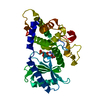
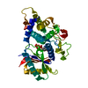
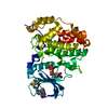
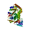
| Entry | Database: PDB / ID: 4nku | ||||||
|---|---|---|---|---|---|---|---|
| Title | Structure of Cid1 in complex with its short product ApU | ||||||
 Components Components |
| ||||||
 Keywords Keywords | TRANSFERASE/RNA / poly(U) polymerase / nucleotidyl tranfer domain / PAP-associated domain / UTP binding / TRANSFERASE-RNA complex | ||||||
| Function / homology |  Function and homology information Function and homology informationpolyuridylation-dependent decapping of nuclear-transcribed mRNA / negative regulation of nuclear-transcribed mRNA poly(A) tail shortening / RNA uridylyltransferase / RNA uridylyltransferase activity / RNA 3'-end processing / polynucleotide adenylyltransferase / poly(A) RNA polymerase activity / UTP binding / magnesium ion binding / RNA binding ...polyuridylation-dependent decapping of nuclear-transcribed mRNA / negative regulation of nuclear-transcribed mRNA poly(A) tail shortening / RNA uridylyltransferase / RNA uridylyltransferase activity / RNA 3'-end processing / polynucleotide adenylyltransferase / poly(A) RNA polymerase activity / UTP binding / magnesium ion binding / RNA binding / ATP binding / cytoplasm / cytosol Similarity search - Function | ||||||
| Biological species |  | ||||||
| Method |  X-RAY DIFFRACTION / SYNCHROTRON / MOLECULAR REPLACEMENT / Resolution: 1.94 Å X-RAY DIFFRACTION / SYNCHROTRON / MOLECULAR REPLACEMENT / Resolution: 1.94 Å | ||||||
 Authors Authors | Munoz-Tello, P. / Gabus, C. / Thore, S. | ||||||
 Citation Citation | Journal: Nucleic Acids Res. / Year: 2014 Title: A critical switch in the enzymatic properties of the Cid1 protein deciphered from its product-bound crystal structure. Authors: Munoz-Tello, P. / Gabus, C. / Thore, S. | ||||||
| History |
|
- Structure visualization
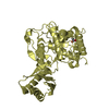
| Structure viewer | Molecule: MolmilJmol/JSmol |
|---|
- Downloads & links
-Download
| PDBx/mmCIF format | 4nku.cif.gz | 150.5 KB | Display | PDBx/mmCIF format |
|---|---|---|---|---|
| PDB format | pdb4nku.ent.gz | 116.2 KB | Display | PDB format |
| PDBx/mmJSON format | 4nku.json.gz | Tree view | PDBx/mmJSON format | |
| Others |  Other downloads Other downloads |
-Validation report
| Arichive directory | https://data.pdbj.org/pub/pdb/validation_reports/nk/4nkuftp://data.pdbj.org/pub/pdb/validation_reports/nk/4nku | HTTPS FTP |
|---|
-Related structure data
| Related structure data |  4nktC 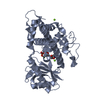 4ep7S C: citing same article ( S: Starting model for refinement |
|---|---|
| Similar structure data |


-Links
 PDBj
PDBj


- Assembly
| Deposited unit | 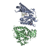
| ||||||||
|---|---|---|---|---|---|---|---|---|---|
| 1 | 
| ||||||||
| 2 | 
| ||||||||
| Unit cell |
|
-Components
| #1: Protein | Mass: 38984.703 Da / Num. of mol.: 2 / Fragment: UNP residues 40-377 / Mutation: D160A Source method: isolated from a genetically manipulated source Source: (gene. exp.) Gene: cid1, SPAC19D5.03 / Plasmid: pET42-based / Production host:  References: UniProt: O13833, Transferases; Transferring phosphorus-containing groups; Nucleotidyltransferases #2: RNA chain | Mass: 590.414 Da / Num. of mol.: 2 / Source method: obtained synthetically #3: Chemical |   Mass: 24.305 Da / Num. of mol.: 3 / Source method: obtained synthetically / Formula: Mg Mass: 24.305 Da / Num. of mol.: 3 / Source method: obtained synthetically / Formula: Mg#4: Chemical |   Mass: 79.904 Da / Num. of mol.: 3 / Source method: obtained synthetically / Formula: Br Mass: 79.904 Da / Num. of mol.: 3 / Source method: obtained synthetically / Formula: Br#5: Water | ChemComp-HOH / |  Mass: 18.015 Da / Num. of mol.: 356 / Source method: isolated from a natural source / Formula: H2O Mass: 18.015 Da / Num. of mol.: 356 / Source method: isolated from a natural source / Formula: H2O |
|---|
-Experimental details
-Experiment
| Experiment | Method: X-RAY DIFFRACTION / Number of used crystals: 1 |
|---|
- Sample preparation
| Crystal | Density Matthews: 2.15 Å3/Da / Density % sol: 42.86 % |
|---|---|
| Crystal grow | Temperature: 291 K / Method: vapor diffusion, sitting drop / pH: 6.1 Details: 0.1 M imidazole/MES, pH 6.1, 20% glycerol, 10% PEG4000, 126 mM halogens (sodium iodide, sodium bromide, sodium fluoride), 10 mM TCEP, VAPOR DIFFUSION, SITTING DROP, temperature 291K |
-Data collection
| Diffraction | Mean temperature: 100 K |
|---|---|
| Diffraction source | Source: SYNCHROTRON / Site: ESRF  / Beamline: ID14-4 / Wavelength: 0.9394 Å / Beamline: ID14-4 / Wavelength: 0.9394 Å |
| Detector | Type: ADSC QUANTUM 315r / Detector: CCD / Date: Jun 4, 2012 |
| Radiation | Monochromator: channel cut Si(111) / Protocol: SINGLE WAVELENGTH / Monochromatic (M) / Laue (L): M / Scattering type: x-ray |
| Radiation wavelength | Wavelength: 0.9394 Å / Relative weight: 1 |
| Reflection | Resolution: 1.94→45.337 Å / Num. all: 49786 / Num. obs: 49569 / % possible obs: 99.5 % / Observed criterion σ(F): 2 / Observed criterion σ(I): 2 / Redundancy: 5 % / Rmerge(I) obs: 0.093 / Net I/σ(I): 13.14 |
| Reflection shell | Highest resolution: 1.94 Å / Redundancy: 5 % / Rmerge(I) obs: 0.086 / Mean I/σ(I) obs: 2.28 / % possible all: 99.2 |
- Processing
| Software |
| |||||||||||||||||||||||||||||||||||||||||||||||||||||||||||||||||||||||||||||||||||||||||||||||||||||||||
|---|---|---|---|---|---|---|---|---|---|---|---|---|---|---|---|---|---|---|---|---|---|---|---|---|---|---|---|---|---|---|---|---|---|---|---|---|---|---|---|---|---|---|---|---|---|---|---|---|---|---|---|---|---|---|---|---|---|---|---|---|---|---|---|---|---|---|---|---|---|---|---|---|---|---|---|---|---|---|---|---|---|---|---|---|---|---|---|---|---|---|---|---|---|---|---|---|---|---|---|---|---|---|---|---|---|---|
| Refinement | Method to determine structure: MOLECULAR REPLACEMENT Starting model: PDB ENTRY 4EP7 Resolution: 1.94→45.337 Å / SU ML: 0.18 / σ(F): 2 / Phase error: 22.77 / Stereochemistry target values: ML
| |||||||||||||||||||||||||||||||||||||||||||||||||||||||||||||||||||||||||||||||||||||||||||||||||||||||||
| Solvent computation | Shrinkage radii: 0.9 Å / VDW probe radii: 1.11 Å / Solvent model: FLAT BULK SOLVENT MODEL | |||||||||||||||||||||||||||||||||||||||||||||||||||||||||||||||||||||||||||||||||||||||||||||||||||||||||
| Refinement step | Cycle: LAST / Resolution: 1.94→45.337 Å
| |||||||||||||||||||||||||||||||||||||||||||||||||||||||||||||||||||||||||||||||||||||||||||||||||||||||||
| Refine LS restraints |
| |||||||||||||||||||||||||||||||||||||||||||||||||||||||||||||||||||||||||||||||||||||||||||||||||||||||||
| LS refinement shell |
|
