regulation of translation / large ribosomal subunit / tRNA binding / rRNA binding / structural constituent of ribosome / translation Similarity search - Function
Ribosomal protein L1/L10, rRNA-binding domain / Ribosomal protein L1/L10, domain II / Ribulose 1,5 Bisphosphate Carboxylase/Oxygenase / Ribosomal protein L1, bacterial-type / Ribosomal protein L1, conserved site / Ribosomal protein L1 signature. / Ribosomal protein L1 / Ribosomal protein L1, 3-layer alpha/beta-sandwich / Ribosomal protein L1-like / Ribosomal protein L1/ribosomal biogenesis protein ...Ribosomal protein L1/L10, rRNA-binding domain / Ribosomal protein L1/L10, domain II / Ribulose 1,5 Bisphosphate Carboxylase/Oxygenase / Ribosomal protein L1, bacterial-type / Ribosomal protein L1, conserved site / Ribosomal protein L1 signature. / Ribosomal protein L1 / Ribosomal protein L1, 3-layer alpha/beta-sandwich / Ribosomal protein L1-like / Ribosomal protein L1/ribosomal biogenesis protein / Ribosomal protein L1p/L10e family / Rossmann fold / 2-Layer Sandwich / 3-Layer(aba) Sandwich / Alpha Beta Similarity search - Domain/homology
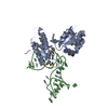
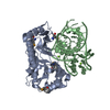
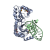
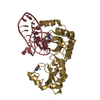
Mass: 12270.358 Da / Num. of mol.: 4 / Source method: obtained synthetically Details: SYNTHESIS OF THE RNA FRAGMENT FROM THE PLASMID PUC18, SEQUENCE FROM CELL-FREE (IN VITRO) SYSTEM WITHOUT LIVING ORGANISM
#2: Protein
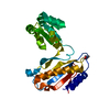
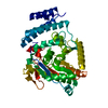
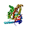
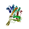
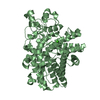
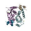
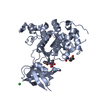
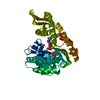
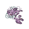
50SribosomalproteinL1
Mass: 24877.188 Da / Num. of mol.: 4 Source method: isolated from a genetically manipulated source Source: (gene. exp.) Thermus thermophilus (bacteria) / Gene: rplA, rpl1 / Plasmid: pACA-L1 / Production host: Escherichia coli (E. coli) / Strain (production host): B834(DE3) / References: UniProt: P27150
In the structure databanks used in Yorodumi, some data are registered as the other names, "COVID-19 virus" and "2019-nCoV". Here are the details of the virus and the list of structure data.
Jan 31, 2019. EMDB accession codes are about to change! (news from PDBe EMDB page)
EMDB accession codes are about to change! (news from PDBe EMDB page)
The allocation of 4 digits for EMDB accession codes will soon come to an end. Whilst these codes will remain in use, new EMDB accession codes will include an additional digit and will expand incrementally as the available range of codes is exhausted. The current 4-digit format prefixed with “EMD-” (i.e. EMD-XXXX) will advance to a 5-digit format (i.e. EMD-XXXXX), and so on. It is currently estimated that the 4-digit codes will be depleted around Spring 2019, at which point the 5-digit format will come into force.
The EM Navigator/Yorodumi systems omit the EMD- prefix.
Related info.:Q: What is EMD? / ID/Accession-code notation in Yorodumi/EM Navigator
Yorodumi is a browser for structure data from EMDB, PDB, SASBDB, etc.
This page is also the successor to EM Navigator detail page, and also detail information page/front-end page for Omokage search.
The word "yorodu" (or yorozu) is an old Japanese word meaning "ten thousand". "mi" (miru) is to see.
Related info.:EMDB / PDB / SASBDB / Comparison of 3 databanks / Yorodumi Search / Aug 31, 2016. New EM Navigator & Yorodumi / Yorodumi Papers / Jmol/JSmol / Function and homology information / Changes in new EM Navigator and Yorodumi
 Movie
Movie Controller


 Open data
 Basic information
 Components
 Keywords
 Function and homology information
 Thermus thermophilus (bacteria)
 X-RAY DIFFRACTION /
  Authors
 Citation
 Structure visualization
 Downloads & links
 Other downloads
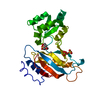


 PDBj
PDBj


 Assembly

 Mass: 39.098 Da / Num. of mol.: 4 / Source method: obtained synthetically / Formula: K
 Mass: 18.015 Da / Num. of mol.: 279 / Source method: isolated from a natural source / Formula: H2O
 Sample preparation
 / Beamline: BW6 / Wavelength: 0.9791 Å
 Processing