 Movie
Movie Controller

[English] 日本語
 Yorodumi
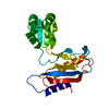
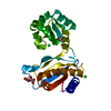
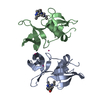
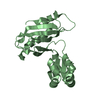
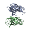
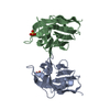
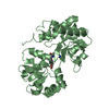
- PDB-1i2a: CRYSTAL STRUCTURE OF L1 RIBOSOMAL PROTEIN FROM METHANOCOCCUS JANN... -
+ Open data

- Basic information
| Entry | Database: PDB / ID: 1i2a | ||||||
|---|---|---|---|---|---|---|---|
| Title | CRYSTAL STRUCTURE OF L1 RIBOSOMAL PROTEIN FROM METHANOCOCCUS JANNASCHII WITH 1.85A RESOLUTION. | ||||||
 Components Components | 50S RIBOSOMAL PROTEIN L1P | ||||||
 Keywords Keywords | RIBOSOME / RIBOSOMAL PROTEIN / PRIMARY RRNA-BINDING PROTEIN / TRANSLATIONAL REPRESSOR | ||||||
| Function / homology |  Function and homology information Function and homology informationregulation of translation / large ribosomal subunit / tRNA binding / rRNA binding / structural constituent of ribosome / translation Similarity search - Function | ||||||
| Biological species |   Methanocaldococcus jannaschii (archaea) Methanocaldococcus jannaschii (archaea) | ||||||
| Method |  X-RAY DIFFRACTION / SYNCHROTRON / MOLECULAR REPLACEMENT / Resolution: 1.85 Å X-RAY DIFFRACTION / SYNCHROTRON / MOLECULAR REPLACEMENT / Resolution: 1.85 Å | ||||||
 Authors Authors | Smolinskaya, Y. / Nikonov, S.V. | ||||||
 Citation Citation | #1: Journal: Structure / Year: 2000Title: Archaeal ribosomal protein L1: the structure provides new insights into RNA binding of the L1 protein family. Authors: Nevskaya, N. / Tischenko, S. / Fedorov, R. / Al-Karadaghi, S. / Liljas, A. / Kraft, A. / Piendl, W. / Garber, M. / Nikonov, S. #2: Journal: Acta Crystallogr.,Sect.D / Year: 2002Title: Structure of ribosomal protein L1 from Methanococcus thermolithotrophicus. Functionally important structural invariants on the L1 surface. Authors: Nevskaya, N. / Tishchenko, S. / Paveliev, M. / Smolinskaya, Y. / Fedorov, R. / Piendl, W. / Nakamura, Y. / Toyoda, T. / Garber, M. / Nikonov, S. | ||||||
| History |
|
- Structure visualization
| Structure viewer | Molecule: MolmilJmol/JSmol |
|---|
- Downloads & links
-Download
| PDBx/mmCIF format | 1i2a.cif.gz | 59 KB | Display | PDBx/mmCIF format |
|---|---|---|---|---|
| PDB format | pdb1i2a.ent.gz | 42.5 KB | Display | PDB format |
| PDBx/mmJSON format | 1i2a.json.gz | Tree view | PDBx/mmJSON format | |
| Others |  Other downloads Other downloads |
-Validation report
| Arichive directory | https://data.pdbj.org/pub/pdb/validation_reports/i2/1i2aftp://data.pdbj.org/pub/pdb/validation_reports/i2/1i2a | HTTPS FTP |
|---|
-Related structure data
| Related structure data |  1cjsSC S: Starting model for refinement C: citing same article ( |
|---|---|
| Similar structure data |


-Links
 PDBj
PDBj


- Assembly
| Deposited unit | 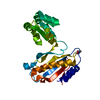
| ||||||||
|---|---|---|---|---|---|---|---|---|---|
| 1 |
| ||||||||
| Unit cell |
|
-Components
| #1: Protein | Mass: 24842.496 Da / Num. of mol.: 1 Source method: isolated from a genetically manipulated source Source: (gene. exp.) Methanocaldococcus jannaschii (archaea)Gene: RPLA / Plasmid: PET11A-MJAL1 / Species (production host): Escherichia coli / Production host:  |
|---|---|
| #2: Chemical | ChemComp-PTL /   Mass: 86.132 Da / Num. of mol.: 1 / Source method: obtained synthetically / Formula: C5H10O Mass: 86.132 Da / Num. of mol.: 1 / Source method: obtained synthetically / Formula: C5H10O |
| #3: Water | ChemComp-HOH /  Mass: 18.015 Da / Num. of mol.: 185 / Source method: isolated from a natural source / Formula: H2O Mass: 18.015 Da / Num. of mol.: 185 / Source method: isolated from a natural source / Formula: H2O |
-Experimental details
-Experiment
| Experiment | Method: X-RAY DIFFRACTION / Number of used crystals: 1 |
|---|
- Sample preparation
| Crystal | Density Matthews: 3.02 Å3/Da / Density % sol: 60 % |
|---|---|
| Crystal grow | Temperature: 301.5 K / Method: vapor diffusion, hanging drop / pH: 7.5 Details: PEG 10000, HEPES-HCl, pH 7.5, VAPOR DIFFUSION, HANGING DROP, temperature 301.5K |
-Data collection
| Diffraction | Mean temperature: 100 K |
|---|---|
| Diffraction source | Source: SYNCHROTRON / Site: MAX II  / Beamline: I711 / Wavelength: 1.009 Å / Beamline: I711 / Wavelength: 1.009 Å |
| Detector | Type: MARRESEARCH / Detector: IMAGE PLATE / Date: Dec 17, 1999 |
| Radiation | Monochromator: Si(111) / Protocol: SINGLE WAVELENGTH / Monochromatic (M) / Laue (L): M / Scattering type: x-ray |
| Radiation wavelength | Wavelength: 1.009 Å / Relative weight: 1 |
| Reflection | Resolution: 1.825→20 Å / Num. all: 188639 / Num. obs: 23155 / % possible obs: 94.8 % / Observed criterion σ(F): 0 / Observed criterion σ(I): 0 / Redundancy: 3.52 % / Biso Wilson estimate: 23.5 Å2 / Rmerge(I) obs: 0.088 / Net I/σ(I): 15 |
| Reflection shell | Resolution: 1.825→2 Å / Redundancy: 3.41 % / Rmerge(I) obs: 0.309 / % possible all: 87.1 |
- Processing
| Software |
| ||||||||||||||||||||||||||||||||||||
|---|---|---|---|---|---|---|---|---|---|---|---|---|---|---|---|---|---|---|---|---|---|---|---|---|---|---|---|---|---|---|---|---|---|---|---|---|---|
| Refinement | Method to determine structure: MOLECULAR REPLACEMENT Starting model: PDB ENTRY 1CJS Resolution: 1.85→19.83 Å / Rfactor Rfree error: 0.008 / Data cutoff high absF: 673607.52 / Data cutoff low absF: 0 / Isotropic thermal model: RESTRAINED / Cross valid method: THROUGHOUT / σ(F): 0 / σ(I): 0
| ||||||||||||||||||||||||||||||||||||
| Solvent computation | Solvent model: FLAT MODEL / Bsol: 56.13 Å2 / ksol: 0.376 e/Å3 | ||||||||||||||||||||||||||||||||||||
| Displacement parameters |
| ||||||||||||||||||||||||||||||||||||
| Refine analyze |
| ||||||||||||||||||||||||||||||||||||
| Refinement step | Cycle: LAST / Resolution: 1.85→19.83 Å
| ||||||||||||||||||||||||||||||||||||
| Refine LS restraints |
| ||||||||||||||||||||||||||||||||||||
| LS refinement shell | Resolution: 1.85→1.97 Å / Rfactor Rfree error: 0.022 / Total num. of bins used: 6
| ||||||||||||||||||||||||||||||||||||
| Xplor file |
|
