Protocol: SINGLE WAVELENGTH / Monochromatic (M) / Laue (L): M / Scattering type: x-ray
Radiation wavelength
Wavelength: 1 Å / Relative weight: 1
Reflection
Resolution: 3.6→70 Å / Num. obs: 4558 / % possible obs: 100 % / Redundancy: 20 % / Net I/σ(I): 28.7
-
Processing
Software
Name
Version
Classification
REFMAC
5.8.0155
refinement
HKL-2000
datareduction
HKL-2000
datascaling
REFMAC
phasing
Refinement
Resolution: 3.64→44.87 Å / Cor.coef. Fo:Fc: 0.901 / Cor.coef. Fo:Fc free: 0.886 / SU B: 45.995 / SU ML: 0.649 / Cross valid method: THROUGHOUT / ESU R Free: 0.781 / Details: HYDROGENS HAVE BEEN ADDED IN THE RIDING POSITIONS
Rfactor
Num. reflection
% reflection
Selection details
Rfree
0.28485
244
5.1 %
RANDOM
Rwork
0.25352
-
-
-
obs
0.25513
4558
99.88 %
-
Solvent computation
Ion probe radii: 0.8 Å / Shrinkage radii: 0.8 Å / VDW probe radii: 1.2 Å
 Movie
Movie Controller


 Open data
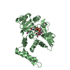
 Basic information
 Components
 Keywords
 Function and homology information
 X-RAY DIFFRACTION /
  Authors
 Citation
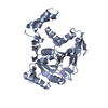
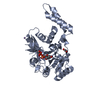
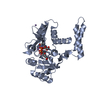
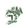
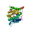
 Structure visualization
 Downloads & links
 Other downloads


 PDBj
PDBj


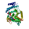
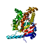
 Assembly
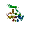

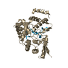
 Type: RNA linking / Mass: 404.161 Da / Num. of mol.: 1 / Source method: obtained synthetically / Formula: C9H14N2O12P2 / Comment: UDP*YM
 Mass: 54.938 Da / Num. of mol.: 1 / Source method: obtained synthetically / Formula: Mn
 Sample preparation
 / Beamline: BL-1A / Wavelength: 1 Å
 Processing