 Movie
Movie Controller

[English] 日本語
 Yorodumi
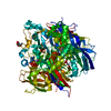
- PDB-5aid: Crystal structure of the Mep2 mutant delta442 from Candida albicans -
+ Open data

- Basic information
| Entry | Database: PDB / ID: 5aid | ||||||
|---|---|---|---|---|---|---|---|
| Title | Crystal structure of the Mep2 mutant delta442 from Candida albicans | ||||||
 Components Components | MEP2 | ||||||
 Keywords Keywords | MEMBRANE PROTEIN / AMMONIUM TRANSPORTER / MEP2 | ||||||
| Function / homology |  Function and homology information Function and homology informationpositive regulation of filamentous growth of a population of unicellular organisms / filamentous growth of a population of unicellular organisms in response to starvation / pseudohyphal growth / nitrogen utilization / ammonium transmembrane transport / ammonium channel activity / filamentous growth / cellular response to nitrogen starvation / cellular response to starvation / plasma membrane Similarity search - Function | ||||||
| Biological species |  CANDIDA ALBICANS (yeast) CANDIDA ALBICANS (yeast) | ||||||
| Method |  X-RAY DIFFRACTION / SYNCHROTRON / MOLECULAR REPLACEMENT / Resolution: 3.4 Å X-RAY DIFFRACTION / SYNCHROTRON / MOLECULAR REPLACEMENT / Resolution: 3.4 Å | ||||||
 Authors Authors | van den Berg, B. / Chembath, A. / Rutherford, J. | ||||||
 Citation Citation | Journal: Nat.Commun. / Year: 2016 Title: Structural Basis for Mep2 Ammonium Transceptor Activation by Phosphorylation. Authors: Van Den Berg, B. / Chembath, A. / Jefferies, D. / Basle, A. / Khalid, S. / Rutherford, J. | ||||||
| History |
|
- Structure visualization
| Structure viewer | Molecule: MolmilJmol/JSmol |
|---|
- Downloads & links
-Download
| PDBx/mmCIF format | 5aid.cif.gz | 86.2 KB | Display | PDBx/mmCIF format |
|---|---|---|---|---|
| PDB format | pdb5aid.ent.gz | 67.3 KB | Display | PDB format |
| PDBx/mmJSON format | 5aid.json.gz | Tree view | PDBx/mmJSON format | |
| Others |  Other downloads Other downloads |
-Validation report
| Arichive directory | https://data.pdbj.org/pub/pdb/validation_reports/ai/5aidftp://data.pdbj.org/pub/pdb/validation_reports/ai/5aid | HTTPS FTP |
|---|
-Related structure data
| Related structure data | 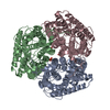 5aexSC 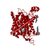 5aezC  5af1C 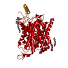 5ah3C 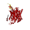 5fufC S: Starting model for refinement C: citing same article ( |
|---|---|
| Similar structure data |


-Links
 PDBj
PDBj- Assembly
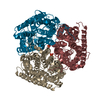
| Deposited unit | 
| ||||||||
|---|---|---|---|---|---|---|---|---|---|
| 1 | 
| ||||||||
| Unit cell |
|
-Components
| #1: Protein | Mass: 48809.207 Da / Num. of mol.: 1 Source method: isolated from a genetically manipulated source Source: (gene. exp.) CANDIDA ALBICANS (yeast) / Plasmid: 83NU / Production host: |
|---|---|
| Sequence details | CONTAINS MUTATIONS R452D S453D |
-Experimental details
-Experiment
| Experiment | Method: X-RAY DIFFRACTION / Number of used crystals: 1 |
|---|
- Sample preparation
| Crystal | Density Matthews: 3.76 Å3/Da / Density % sol: 67 % / Description: NONE |
|---|---|
| Crystal grow | pH: 6 Details: 24% PEG400, 0.05 M SODIUM ACETATE, 0.05 M MAGNESIUM ACETATE PH 6.1 |
-Data collection
| Diffraction | Mean temperature: 100 K |
|---|---|
| Diffraction source | Source: SYNCHROTRON / Site: Diamond  / Beamline: I04-1 / Wavelength: 0.92 / Beamline: I04-1 / Wavelength: 0.92 |
| Detector | Type: DEXTRIS / Detector: PIXEL / Date: Aug 4, 2014 |
| Radiation | Protocol: SINGLE WAVELENGTH / Monochromatic (M) / Laue (L): M / Scattering type: x-ray |
| Radiation wavelength | Wavelength: 0.92 Å / Relative weight: 1 |
| Reflection | Resolution: 3.4→96.65 Å / Num. obs: 10453 / % possible obs: 100 % / Observed criterion σ(I): -3 / Redundancy: 9.3 % / Biso Wilson estimate: 132.97 Å2 / Rmerge(I) obs: 0.1 / Net I/σ(I): 14.6 |
| Reflection shell | Resolution: 3.4→3.67 Å / Redundancy: 9.7 % / Rmerge(I) obs: 1.17 / Mean I/σ(I) obs: 2.5 / % possible all: 100 |
- Processing
| Software |
| |||||||||||||||||||||||||||||||||||||||||||||||||||||||||||||||
|---|---|---|---|---|---|---|---|---|---|---|---|---|---|---|---|---|---|---|---|---|---|---|---|---|---|---|---|---|---|---|---|---|---|---|---|---|---|---|---|---|---|---|---|---|---|---|---|---|---|---|---|---|---|---|---|---|---|---|---|---|---|---|---|---|
| Refinement | Method to determine structure: MOLECULAR REPLACEMENT Starting model: PDB ENTRY 5AEX Resolution: 3.4→96.655 Å / SU ML: 0.44 / σ(F): 1.33 / Phase error: 34.85 / Stereochemistry target values: ML
| |||||||||||||||||||||||||||||||||||||||||||||||||||||||||||||||
| Solvent computation | Shrinkage radii: 0.9 Å / VDW probe radii: 1.11 Å / Solvent model: FLAT BULK SOLVENT MODEL | |||||||||||||||||||||||||||||||||||||||||||||||||||||||||||||||
| Refinement step | Cycle: LAST / Resolution: 3.4→96.655 Å
| |||||||||||||||||||||||||||||||||||||||||||||||||||||||||||||||
| Refine LS restraints |
| |||||||||||||||||||||||||||||||||||||||||||||||||||||||||||||||
| LS refinement shell |
|
