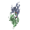
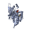
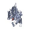
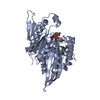
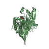
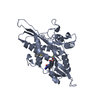
Kinesin-likeproteinKIF11 / Kinesin-related motor protein Eg5 / Kinesin-like spindle protein HKSP / Thyroid receptor- ...Kinesin-related motor protein Eg5 / Kinesin-like spindle protein HKSP / Thyroid receptor-interacting protein 5 / TRIP-5 / Kinesin-like protein 1
Mass: 41168.738 Da / Num. of mol.: 1 Fragment: motor domain of human kinesin Eg5 (residues 1-369) Source method: isolated from a genetically manipulated source Source: (gene. exp.) Homo sapiens (human) / Gene: EG5, KIF11, KNSL1, TRIP5 / Plasmid: pET28a / Production host: Escherichia coli (E. coli) / Strain (production host): BL21(DE3) / References: UniProt: P52732
Mass: 18.015 Da / Num. of mol.: 74 / Source method: isolated from a natural source / Formula: H2O
-
Experimental details
-
Experiment
Experiment
Method: X-RAY DIFFRACTION / Number of used crystals: 1
-
Sample preparation
Crystal
Density Matthews: 3.99 Å3/Da / Density % sol: 69.15 %
Crystal grow
Temperature: 277 K / Method: vapor diffusion, sitting drop / pH: 6 Details: 250 mM NH4SO4, 100 mM MES pH 6.0, 25% PEG 3350, 5-15% glycerol, 10 mM trimethylamine, VAPOR DIFFUSION, SITTING DROP, temperature 277K
-
Data collection
Diffraction
Mean temperature: 100 K
Diffraction source
Source: ROTATING ANODE / Type: BRUKER AXS MICROSTAR / Wavelength: 1.5418 Å
Detector
Type: Bruker Platinum 135 / Detector: CCD / Date: Aug 13, 2008
Radiation
Monochromator: Ni Filter / Protocol: SINGLE WAVELENGTH / Monochromatic (M) / Laue (L): M / Scattering type: x-ray
In the structure databanks used in Yorodumi, some data are registered as the other names, "COVID-19 virus" and "2019-nCoV". Here are the details of the virus and the list of structure data.
Jan 31, 2019. EMDB accession codes are about to change! (news from PDBe EMDB page)
EMDB accession codes are about to change! (news from PDBe EMDB page)
The allocation of 4 digits for EMDB accession codes will soon come to an end. Whilst these codes will remain in use, new EMDB accession codes will include an additional digit and will expand incrementally as the available range of codes is exhausted. The current 4-digit format prefixed with “EMD-” (i.e. EMD-XXXX) will advance to a 5-digit format (i.e. EMD-XXXXX), and so on. It is currently estimated that the 4-digit codes will be depleted around Spring 2019, at which point the 5-digit format will come into force.
The EM Navigator/Yorodumi systems omit the EMD- prefix.
Related info.:Q: What is EMD? / ID/Accession-code notation in Yorodumi/EM Navigator
Yorodumi is a browser for structure data from EMDB, PDB, SASBDB, etc.
This page is also the successor to EM Navigator detail page, and also detail information page/front-end page for Omokage search.
The word "yorodu" (or yorozu) is an old Japanese word meaning "ten thousand". "mi" (miru) is to see.
Related info.:EMDB / PDB / SASBDB / Comparison of 3 databanks / Yorodumi Search / Aug 31, 2016. New EM Navigator & Yorodumi / Yorodumi Papers / Jmol/JSmol / Function and homology information / Changes in new EM Navigator and Yorodumi
 Movie
Movie Controller


 Open data
 Basic information
 Components
 Keywords
 Function and homology information
 Homo sapiens (human)
 X-RAY DIFFRACTION /
  Authors
 Citation
 Structure visualization
 Downloads & links
 Other downloads


 PDBj
PDBj


 Assembly
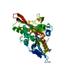


 Mass: 427.201 Da / Num. of mol.: 1 / Source method: obtained synthetically / Formula: C10H15N5O10P2 / Comment: ADP, energy-carrying molecule*YM
 Type: L-peptide linking / Mass: 363.473 Da / Num. of mol.: 1 / Source method: obtained synthetically / Formula: C22H21NO2S
 Mass: 59.110 Da / Num. of mol.: 1 / Source method: obtained synthetically / Formula: C3H9N
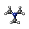
 Mass: 24.305 Da / Num. of mol.: 1 / Source method: obtained synthetically / Formula: Mg
 Sample preparation
 Processing