 Movie
Movie Controller

[English] 日本語
 Yorodumi
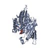
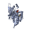
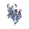
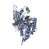
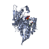
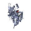
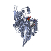
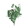
- PDB-1q0b: Crystal structure of the motor protein KSP in complex with ADP an... -
+ Open data

- Basic information
| Entry | Database: PDB / ID: 1q0b | ||||||
|---|---|---|---|---|---|---|---|
| Title | Crystal structure of the motor protein KSP in complex with ADP and monastrol | ||||||
 Components Components | Kinesin-like protein KIF11 | ||||||
 Keywords Keywords | CELL CYCLE / motor protein / monastrol | ||||||
| Function / homology |  Function and homology information Function and homology informationspindle elongation / regulation of mitotic centrosome separation / plus-end-directed microtubule motor activity / Kinesins / spindle organization / microtubule motor activity / kinesin complex / COPI-dependent Golgi-to-ER retrograde traffic / microtubule-based movement / mitotic spindle assembly ...spindle elongation / regulation of mitotic centrosome separation / plus-end-directed microtubule motor activity / Kinesins / spindle organization / microtubule motor activity / kinesin complex / COPI-dependent Golgi-to-ER retrograde traffic / microtubule-based movement / mitotic spindle assembly / MHC class II antigen presentation / mitotic spindle organization / spindle / spindle pole / mitotic spindle / mitotic cell cycle / microtubule binding / microtubule / cell division / protein kinase binding / protein-containing complex / ATP binding / membrane / nucleus / cytosol Similarity search - Function | ||||||
| Biological species |  Homo sapiens (human) Homo sapiens (human) | ||||||
| Method |  X-RAY DIFFRACTION / SYNCHROTRON / MOLECULAR REPLACEMENT / Resolution: 1.9 Å X-RAY DIFFRACTION / SYNCHROTRON / MOLECULAR REPLACEMENT / Resolution: 1.9 Å | ||||||
 Authors Authors | Yan, Y. / Sardana, V. / Xu, B. / Halczenko, W. / Homnick, C. / Buser, C.A. / Hartman, G.D. / Huber, H.E. / Kuo, L.C. | ||||||
 Citation Citation | Journal: J.Mol.Biol. / Year: 2004 Title: Inhibition of a mitotic motor protein: where, how, and conformational consequences Authors: Yan, Y. / Sardana, V. / Xu, B. / Homnick, C. / Halczenko, W. / Buser, C.A. / Schaber, M. / Hartman, G.D. / Huber, H.E. / Kuo, L.C. | ||||||
| History |
|
- Structure visualization
| Structure viewer | Molecule: MolmilJmol/JSmol |
|---|
- Downloads & links
-Download
| PDBx/mmCIF format | 1q0b.cif.gz | 157.9 KB | Display | PDBx/mmCIF format |
|---|---|---|---|---|
| PDB format | pdb1q0b.ent.gz | 122.3 KB | Display | PDB format |
| PDBx/mmJSON format | 1q0b.json.gz | Tree view | PDBx/mmJSON format | |
| Others |  Other downloads Other downloads |
-Validation report
| Arichive directory | https://data.pdbj.org/pub/pdb/validation_reports/q0/1q0bftp://data.pdbj.org/pub/pdb/validation_reports/q0/1q0b | HTTPS FTP |
|---|
-Related structure data
| Related structure data | 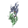 1ii6S S: Starting model for refinement |
|---|---|
| Similar structure data |


-Links
 PDBj
PDBj


- Assembly
| Deposited unit | 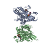
| ||||||||
|---|---|---|---|---|---|---|---|---|---|
| 1 | 
| ||||||||
| 2 | 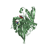
| ||||||||
| Unit cell |
|
-Components
| #1: Protein | Mass: 40924.387 Da / Num. of mol.: 2 / Fragment: residues 2-368 Source method: isolated from a genetically manipulated source Source: (gene. exp.) Homo sapiens (human) / Gene: KIF11 OR KNSL1 OR EG5 / Plasmid: pRSETa / Species (production host): Escherichia coli / Production host:  #2: Chemical |   Mass: 24.305 Da / Num. of mol.: 2 / Source method: obtained synthetically / Formula: Mg Mass: 24.305 Da / Num. of mol.: 2 / Source method: obtained synthetically / Formula: Mg#3: Chemical |   Mass: 427.201 Da / Num. of mol.: 2 / Source method: obtained synthetically / Formula: C10H15N5O10P2 / Comment: ADP, energy-carrying molecule*YM Mass: 427.201 Da / Num. of mol.: 2 / Source method: obtained synthetically / Formula: C10H15N5O10P2 / Comment: ADP, energy-carrying molecule*YM#4: Chemical |   Mass: 292.353 Da / Num. of mol.: 2 / Source method: obtained synthetically / Formula: C14H16N2O3S / Comment: inhibitor*YM Mass: 292.353 Da / Num. of mol.: 2 / Source method: obtained synthetically / Formula: C14H16N2O3S / Comment: inhibitor*YM#5: Water | ChemComp-HOH / |  Mass: 18.015 Da / Num. of mol.: 500 / Source method: isolated from a natural source / Formula: H2O Mass: 18.015 Da / Num. of mol.: 500 / Source method: isolated from a natural source / Formula: H2O |
|---|
-Experimental details
-Experiment
| Experiment | Method: X-RAY DIFFRACTION / Number of used crystals: 1 |
|---|
- Sample preparation
| Crystal | Density Matthews: 2.68 Å3/Da / Density % sol: 54.07 % | ||||||||||||||||||||||||||||||||||||||||||||||||||||||||||||||||||||||
|---|---|---|---|---|---|---|---|---|---|---|---|---|---|---|---|---|---|---|---|---|---|---|---|---|---|---|---|---|---|---|---|---|---|---|---|---|---|---|---|---|---|---|---|---|---|---|---|---|---|---|---|---|---|---|---|---|---|---|---|---|---|---|---|---|---|---|---|---|---|---|---|
| Crystal grow | Temperature: 277 K / Method: vapor diffusion, hanging drop / pH: 8 Details: PEG 3350, potassium phosphate, pH 8, VAPOR DIFFUSION, HANGING DROP, temperature 277K | ||||||||||||||||||||||||||||||||||||||||||||||||||||||||||||||||||||||
| Crystal grow | *PLUS pH: 7 / Method: vapor diffusion, hanging drop / Details: used macroseeding | ||||||||||||||||||||||||||||||||||||||||||||||||||||||||||||||||||||||
| Components of the solutions | *PLUS
|
-Data collection
| Diffraction | Mean temperature: 100 K |
|---|---|
| Diffraction source | Source: SYNCHROTRON / Site: APS  / Beamline: 17-ID / Wavelength: 1 Å / Beamline: 17-ID / Wavelength: 1 Å |
| Detector | Type: ADSC QUANTUM 4 / Detector: CCD / Date: Jul 16, 2002 |
| Radiation | Protocol: SINGLE WAVELENGTH / Monochromatic (M) / Laue (L): M / Scattering type: x-ray |
| Radiation wavelength | Wavelength: 1 Å / Relative weight: 1 |
| Reflection | Resolution: 1.9→20 Å / Num. all: 71000 / Num. obs: 70259 / % possible obs: 99 % / Observed criterion σ(F): 0 / Observed criterion σ(I): 0 / Rmerge(I) obs: 0.067 / Net I/σ(I): 26 |
| Reflection shell | Resolution: 1.9→1.96 Å / Rmerge(I) obs: 0.24 / Mean I/σ(I) obs: 4.5 / % possible all: 97 |
| Reflection | *PLUS Lowest resolution: 20 Å |
| Reflection shell | *PLUS % possible obs: 97 % |
- Processing
| Software |
| |||||||||||||||||||||||||
|---|---|---|---|---|---|---|---|---|---|---|---|---|---|---|---|---|---|---|---|---|---|---|---|---|---|---|
| Refinement | Method to determine structure: MOLECULAR REPLACEMENT Starting model: PDB entry 1II6 Resolution: 1.9→20 Å / Data cutoff low absF: 0 / Isotropic thermal model: isotropic / σ(F): 0 / σ(I): 0 / Stereochemistry target values: Engh & Huber
| |||||||||||||||||||||||||
| Refinement step | Cycle: LAST / Resolution: 1.9→20 Å
| |||||||||||||||||||||||||
| Refine LS restraints |
| |||||||||||||||||||||||||
| Refinement | *PLUS Lowest resolution: 20 Å / % reflection Rfree: 10 % | |||||||||||||||||||||||||
| Solvent computation | *PLUS | |||||||||||||||||||||||||
| Displacement parameters | *PLUS |
