 Movie
Movie Controller

[English] 日本語
 Yorodumi
- PDB-2nqj: Crystal structure of Escherichia coli endonuclease IV (Endo IV) E... -
+ Open data

- Basic information
| Entry | Database: PDB / ID: 2nqj | ||||||
|---|---|---|---|---|---|---|---|
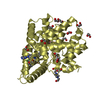
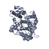
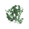
| Title | Crystal structure of Escherichia coli endonuclease IV (Endo IV) E261Q mutant bound to damaged DNA | ||||||
 Components Components |
| ||||||
 Keywords Keywords | HYDROLASE / Tim_barrel / trinuclear zinc site | ||||||
| Function / homology |  Function and homology information Function and homology informationdeoxyribonuclease IV / deoxyribonuclease IV (phage-T4-induced) activity / phosphoric diester hydrolase activity / 3'-5'-DNA exonuclease activity / phosphatase activity / DNA-(apurinic or apyrimidinic site) endonuclease activity / base-excision repair / DNA repair / DNA binding / zinc ion binding / cytosol Similarity search - Function | ||||||
| Biological species |  | ||||||
| Method |  X-RAY DIFFRACTION / SYNCHROTRON / MOLECULAR REPLACEMENT / Resolution: 2.45 Å X-RAY DIFFRACTION / SYNCHROTRON / MOLECULAR REPLACEMENT / Resolution: 2.45 Å | ||||||
 Authors Authors | Garcin-Hosfield, E.D. / Hosfield, D.J. / Tainer, J.A. | ||||||
 Citation Citation | Journal: Nat.Struct.Mol.Biol. / Year: 2008 Title: DNA apurinic-apyrimidinic site binding and excision by endonuclease IV. Authors: Garcin, E.D. / Hosfield, D.J. / Desai, S.A. / Haas, B.J. / Bjoras, M. / Cunningham, R.P. / Tainer, J.A. | ||||||
| History |
| ||||||
| Remark 300 | BIOMOLECULE: 1, 2 THIS ENTRY CONTAINS THE CRYSTALLOGRAPHIC ASYMMETRIC UNIT WHICH CONSISTS OF 4 ... BIOMOLECULE: 1, 2 THIS ENTRY CONTAINS THE CRYSTALLOGRAPHIC ASYMMETRIC UNIT WHICH CONSISTS OF 4 CHAIN(S). SEE REMARK 350 FOR INFORMATION ON GENERATING THE BIOLOGICAL MOLECULE(S). ALTHOUGH CHAIN B COULD BE MOVED BY A SYMMETRY OPERATION, THE COMPLEX FORMED BY CHAIN B WITH CHAIN C AND D IS NOT BIOLOGICALLY RELEVANT. |
- Structure visualization
| Structure viewer | Molecule: MolmilJmol/JSmol |
|---|
- Downloads & links
-Download
| PDBx/mmCIF format | 2nqj.cif.gz | 146.1 KB | Display | PDBx/mmCIF format |
|---|---|---|---|---|
| PDB format | pdb2nqj.ent.gz | 109.6 KB | Display | PDB format |
| PDBx/mmJSON format | 2nqj.json.gz | Tree view | PDBx/mmJSON format | |
| Others |  Other downloads Other downloads |
-Validation report
| Arichive directory | https://data.pdbj.org/pub/pdb/validation_reports/nq/2nqjftp://data.pdbj.org/pub/pdb/validation_reports/nq/2nqj | HTTPS FTP |
|---|
-Related structure data
| Related structure data | 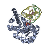 2nq9C  2nqhC  1qtwS C: citing same article ( S: Starting model for refinement |
|---|---|
| Similar structure data |


-Links
 PDBj
PDBj


- Assembly
| Deposited unit | 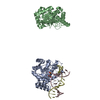
| ||||||||
|---|---|---|---|---|---|---|---|---|---|
| 1 | 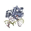
| ||||||||
| 2 | 
| ||||||||
| Unit cell |
|
-Components
| #1: DNA chain | Mass: 4446.864 Da / Num. of mol.: 1 / Source method: obtained synthetically | ||||
|---|---|---|---|---|---|
| #2: DNA chain | Mass: 4626.984 Da / Num. of mol.: 1 / Source method: obtained synthetically | ||||
| #3: Protein | Mass: 31517.512 Da / Num. of mol.: 2 / Mutation: E261Q Source method: isolated from a genetically manipulated source Source: (gene. exp.) #4: Chemical | ChemComp-ZN /   Mass: 65.409 Da / Num. of mol.: 4 / Source method: obtained synthetically / Formula: Zn Mass: 65.409 Da / Num. of mol.: 4 / Source method: obtained synthetically / Formula: Zn#5: Water | ChemComp-HOH / |  Mass: 18.015 Da / Num. of mol.: 325 / Source method: isolated from a natural source / Formula: H2O Mass: 18.015 Da / Num. of mol.: 325 / Source method: isolated from a natural source / Formula: H2O |
-Experimental details
-Experiment
| Experiment | Method: X-RAY DIFFRACTION / Number of used crystals: 1 |
|---|
- Sample preparation
| Crystal | Density Matthews: 2.45 Å3/Da / Density % sol: 49.79 % | ||||||||||||||||||||||||||||
|---|---|---|---|---|---|---|---|---|---|---|---|---|---|---|---|---|---|---|---|---|---|---|---|---|---|---|---|---|---|
| Crystal grow | Temperature: 298 K / Method: vapor diffusion, hanging drop / pH: 7 Details: 20% MPEG-2000, 0.1M HEPES pH 7, 0.4M sodium acetate, pH 7.0, VAPOR DIFFUSION, HANGING DROP, temperature 298K | ||||||||||||||||||||||||||||
| Components of the solutions |
|
-Data collection
| Diffraction | Mean temperature: 100 K |
|---|---|
| Diffraction source | Source: SYNCHROTRON / Site: SSRL  / Beamline: BL9-1 / Wavelength: 0.98 Å / Beamline: BL9-1 / Wavelength: 0.98 Å |
| Detector | Type: MARRESEARCH / Detector: IMAGE PLATE / Date: Oct 10, 1998 |
| Radiation | Protocol: SINGLE WAVELENGTH / Monochromatic (M) / Laue (L): M / Scattering type: x-ray |
| Radiation wavelength | Wavelength: 0.98 Å / Relative weight: 1 |
| Reflection | Resolution: 2.45→20 Å / Num. obs: 24943 / % possible obs: 93 % / Rsym value: 0.095 / Net I/σ(I): 14.6 |
- Processing
| Software |
| ||||||||||||||||||||||||
|---|---|---|---|---|---|---|---|---|---|---|---|---|---|---|---|---|---|---|---|---|---|---|---|---|---|
| Refinement | Method to determine structure: MOLECULAR REPLACEMENT Starting model: 1QTW Resolution: 2.45→20 Å / σ(F): 0
| ||||||||||||||||||||||||
| Solvent computation | Bsol: 50.958 Å2 | ||||||||||||||||||||||||
| Displacement parameters | Biso mean: 36.078 Å2
| ||||||||||||||||||||||||
| Refinement step | Cycle: LAST / Resolution: 2.45→20 Å
| ||||||||||||||||||||||||
| Refine LS restraints |
| ||||||||||||||||||||||||
| Xplor file |
|
