 Movie
Movie Controller

[English] 日本語
 Yorodumi
- PDB-4in9: Structure of karilysin MMP-like catalytic domain in complex with ... -
+ Open data

- Basic information
| Entry | Database: PDB / ID: 4in9 | ||||||
|---|---|---|---|---|---|---|---|
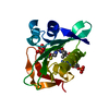
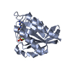
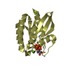
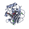
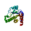
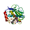
| Title | Structure of karilysin MMP-like catalytic domain in complex with inhibitory tetrapeptide SWFP | ||||||
 Components Components |
| ||||||
 Keywords Keywords | HYDROLASE / matrixin / metallopeptidase / metalloprotease / hydrolytic enzyme | ||||||
| Function / homology |  Function and homology information Function and homology informationHydrolases; Acting on peptide bonds (peptidases); Metalloendopeptidases / collagen catabolic process / extracellular matrix organization / metalloendopeptidase activity / extracellular matrix / proteolysis / extracellular region / zinc ion binding Similarity search - Function | ||||||
| Biological species |  Tannerella forsythia (bacteria) Tannerella forsythia (bacteria)synthetic construct (others) | ||||||
| Method |  X-RAY DIFFRACTION / SYNCHROTRON / FOURIER SYNTHESIS / Resolution: 1.55 Å X-RAY DIFFRACTION / SYNCHROTRON / FOURIER SYNTHESIS / Resolution: 1.55 Å | ||||||
 Authors Authors | Guevara, T. / Ksiazek, M. / Skottrup, P.D. / Cerda-Costa, N. / Trillo-Muyo, S. / de Diego, I. / Riise, E. / Potempa, J. / Gomis-Ruth, F.X. | ||||||
 Citation Citation | Journal: Acta Crystallogr.,Sect.F / Year: 2013 Title: Structure of the catalytic domain of the Tannerella forsythia matrix metallopeptidase karilysin in complex with a tetrapeptidic inhibitor. Authors: Guevara, T. / Ksiazek, M. / Skottrup, P.D. / Cerda-Costa, N. / Trillo-Muyo, S. / de Diego, I. / Riise, E. / Potempa, J. / Gomis-Ruth, F.X. #1: Journal: Mol.Microbiol. / Year: 2011Title: The structure of the catalytic domain of Tannerella forsythia karilysin reveals it is a bacterial xenologue of animal matrix metalloproteinases. Authors: Cerda-Costa, N. / Guevara, T. / Karim, A.Y. / Ksiazek, M. / Nguyen, K.A. / Arolas, J.L. / Potempa, J. / Gomis-Ruth, F.X. | ||||||
| History |
|
- Structure visualization
| Structure viewer | Molecule: MolmilJmol/JSmol |
|---|
- Downloads & links
-Download
| PDBx/mmCIF format | 4in9.cif.gz | 88.2 KB | Display | PDBx/mmCIF format |
|---|---|---|---|---|
| PDB format | pdb4in9.ent.gz | 64.9 KB | Display | PDB format |
| PDBx/mmJSON format | 4in9.json.gz | Tree view | PDBx/mmJSON format | |
| Others |  Other downloads Other downloads |
-Validation report
| Arichive directory | https://data.pdbj.org/pub/pdb/validation_reports/in/4in9ftp://data.pdbj.org/pub/pdb/validation_reports/in/4in9 | HTTPS FTP |
|---|
-Related structure data
| Related structure data |  2xs4S S: Starting model for refinement |
|---|---|
| Similar structure data |


-Links
 PDBj
PDBj


- Assembly
| Deposited unit | 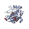
| ||||||||
|---|---|---|---|---|---|---|---|---|---|
| 1 |
| ||||||||
| Unit cell |
|
-Components
-Protein / Protein/peptide , 2 types, 2 molecules AB
| #1: Protein | Mass: 18703.615 Da / Num. of mol.: 1 / Fragment: UNP residues 35-200 Source method: isolated from a genetically manipulated source Source: (gene. exp.) Tannerella forsythia (bacteria) / Production host: |
|---|---|
| #2: Protein/peptide | Mass: 535.591 Da / Num. of mol.: 1 / Source method: obtained synthetically Details: Synthetic peptide arising from phage-display screening Source: (synth.) synthetic construct (others) |
-Non-polymers , 5 types, 217 molecules 


| #3: Chemical |  Mass: 65.409 Da / Num. of mol.: 2 / Source method: obtained synthetically / Formula: Zn Mass: 65.409 Da / Num. of mol.: 2 / Source method: obtained synthetically / Formula: Zn#4: Chemical | ChemComp-K / |  Mass: 39.098 Da / Num. of mol.: 1 / Source method: obtained synthetically / Formula: K Mass: 39.098 Da / Num. of mol.: 1 / Source method: obtained synthetically / Formula: K#5: Chemical | ChemComp-NA / |  Mass: 22.990 Da / Num. of mol.: 1 / Source method: obtained synthetically / Formula: Na Mass: 22.990 Da / Num. of mol.: 1 / Source method: obtained synthetically / Formula: Na#6: Chemical | ChemComp-GOL /  Mass: 92.094 Da / Num. of mol.: 5 / Source method: obtained synthetically / Formula: C3H8O3 Mass: 92.094 Da / Num. of mol.: 5 / Source method: obtained synthetically / Formula: C3H8O3#7: Water | ChemComp-HOH / | Mass: 18.015 Da / Num. of mol.: 208 / Source method: isolated from a natural source / Formula: H2O |
|---|
-Experimental details
-Experiment
| Experiment | Method: X-RAY DIFFRACTION / Number of used crystals: 1 |
|---|
- Sample preparation
| Crystal | Density Matthews: 2.38 Å3/Da / Density % sol: 48.29 % |
|---|---|
| Crystal grow | Temperature: 293 K / Method: vapor diffusion, sitting drop / pH: 8 Details: Best crystals were obtained at 20 degrees from 1:1 microL drops with complex solution (8mg/mL in 5mM Tris HCl pH8, 5mM calcium chloride, 0.02% sodium azide) and 0.4M sodium/potassium ...Details: Best crystals were obtained at 20 degrees from 1:1 microL drops with complex solution (8mg/mL in 5mM Tris HCl pH8, 5mM calcium chloride, 0.02% sodium azide) and 0.4M sodium/potassium tartrate as reservoir solution. Crystals were cryo-protected by immersion in cryo-solution (0.32M sodium/potassium tartrate, 25%[v/v] glycerol), VAPOR DIFFUSION, SITTING DROP, temperature 293K |
-Data collection
| Diffraction | Mean temperature: 100 K |
|---|---|
| Diffraction source | Source: SYNCHROTRON / Site: ESRF  / Beamline: ID14-4 / Wavelength: 0.9393 Å / Beamline: ID14-4 / Wavelength: 0.9393 Å |
| Detector | Type: ADSC QUANTUM 315r / Detector: CCD / Date: Jul 31, 2011 |
| Radiation | Monochromator: channel cut ESRF monochromator / Protocol: SINGLE WAVELENGTH / Monochromatic (M) / Laue (L): M / Scattering type: x-ray |
| Radiation wavelength | Wavelength: 0.9393 Å / Relative weight: 1 |
| Reflection | Resolution: 1.55→42.9 Å / Num. all: 26370 / Num. obs: 26344 / % possible obs: 99.9 % / Observed criterion σ(F): 0 / Observed criterion σ(I): 2 / Redundancy: 7 % / Biso Wilson estimate: 21.2 Å2 / Rmerge(I) obs: 0.081 / Net I/σ(I): 18.4 |
| Reflection shell | Resolution: 1.55→1.59 Å / Redundancy: 4.3 % / Rmerge(I) obs: 0.749 / Mean I/σ(I) obs: 2.1 / % possible all: 98.7 |
- Processing
| Software |
| ||||||||||||||||||||||||||||||||||||||||||||||||||||||||||||||||||||||||||||||
|---|---|---|---|---|---|---|---|---|---|---|---|---|---|---|---|---|---|---|---|---|---|---|---|---|---|---|---|---|---|---|---|---|---|---|---|---|---|---|---|---|---|---|---|---|---|---|---|---|---|---|---|---|---|---|---|---|---|---|---|---|---|---|---|---|---|---|---|---|---|---|---|---|---|---|---|---|---|---|---|
| Refinement | Method to determine structure: FOURIER SYNTHESIS Starting model: 2XS4 Resolution: 1.55→42.87 Å / Cor.coef. Fo:Fc: 0.9679 / Cor.coef. Fo:Fc free: 0.9567 / SU R Cruickshank DPI: 0.064 / Cross valid method: THROUGHOUT / σ(F): 0 / Stereochemistry target values: Engh & Huber
| ||||||||||||||||||||||||||||||||||||||||||||||||||||||||||||||||||||||||||||||
| Displacement parameters | Biso mean: 17.58 Å2
| ||||||||||||||||||||||||||||||||||||||||||||||||||||||||||||||||||||||||||||||
| Refine analyze | Luzzati coordinate error obs: 0.137 Å | ||||||||||||||||||||||||||||||||||||||||||||||||||||||||||||||||||||||||||||||
| Refinement step | Cycle: LAST / Resolution: 1.55→42.87 Å
| ||||||||||||||||||||||||||||||||||||||||||||||||||||||||||||||||||||||||||||||
| Refine LS restraints |
| ||||||||||||||||||||||||||||||||||||||||||||||||||||||||||||||||||||||||||||||
| LS refinement shell | Resolution: 1.55→1.61 Å / Total num. of bins used: 13
| ||||||||||||||||||||||||||||||||||||||||||||||||||||||||||||||||||||||||||||||
| Refinement TLS params. | Method: refined / Refine-ID: X-RAY DIFFRACTION
| ||||||||||||||||||||||||||||||||||||||||||||||||||||||||||||||||||||||||||||||
| Refinement TLS group |
|
