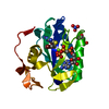
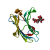
small GTPase-mediated signal transduction / TOR signaling / : / endomembrane system / GDP binding / Hydrolases; Acting on acid anhydrides; Acting on GTP to facilitate cellular and subcellular movement / GTPase activity / GTP binding / metal ion binding / plasma membrane / cytoplasm Similarity search - Function
Small GTPase, Ras-type / Small GTPase Ras domain profile. / Rho (Ras homology) subfamily of Ras-like small GTPases / Ras subfamily of RAS small GTPases / Small GTPase / Ras family / Rab subfamily of small GTPases / Small GTP-binding protein domain / P-loop containing nucleotide triphosphate hydrolases / Rossmann fold ...Small GTPase, Ras-type / Small GTPase Ras domain profile. / Rho (Ras homology) subfamily of Ras-like small GTPases / Ras subfamily of RAS small GTPases / Small GTPase / Ras family / Rab subfamily of small GTPases / Small GTP-binding protein domain / P-loop containing nucleotide triphosphate hydrolases / Rossmann fold / P-loop containing nucleoside triphosphate hydrolase / 3-Layer(aba) Sandwich / Alpha Beta Similarity search - Domain/homology
Resolution: 2.301→20 Å / Cor.coef. Fo:Fc: 0.944 / Cor.coef. Fo:Fc free: 0.911 / WRfactor Rfree: 0.242 / WRfactor Rwork: 0.192 / SU B: 7.291 / SU ML: 0.177 / Cross valid method: THROUGHOUT / σ(F): 0 / ESU R Free: 0.245 / Stereochemistry target values: MAXIMUM LIKELIHOOD Details: HYDROGENS HAVE BEEN ADDED IN THE RIDING POSITIONS. U VALUES REFINED INDIVIDUALLY. Programs chainsaw, coot and the molprobity server were also used during refinement.
Rfactor
Num. reflection
% reflection
Selection details
Rfree
0.271
418
4.766 %
RANDOM
Rwork
0.212
-
-
-
obs
0.215
8770
99.523 %
-
Solvent computation
Ion probe radii: 0.8 Å / Shrinkage radii: 0.8 Å / VDW probe radii: 1.4 Å / Solvent model: MASK BULK SOLVENT
Displacement parameters
Biso mean: 32.946 Å2
Baniso -1
Baniso -2
Baniso -3
1-
-1.669 Å2
0 Å2
0 Å2
2-
-
3.182 Å2
0 Å2
3-
-
-
-1.513 Å2
Refinement step
Cycle: LAST / Resolution: 2.301→20 Å
Protein
Nucleic acid
Ligand
Solvent
Total
Num. atoms
1195
0
33
13
1241
Refine LS restraints
Refine-ID
Type
Dev ideal
Dev ideal target
Number
X-RAY DIFFRACTION
r_bond_refined_d
0.015
0.022
1252
X-RAY DIFFRACTION
r_bond_other_d
0.002
0.02
795
X-RAY DIFFRACTION
r_angle_refined_deg
1.399
1.982
1705
X-RAY DIFFRACTION
r_angle_other_deg
0.86
3
1946
X-RAY DIFFRACTION
r_dihedral_angle_1_deg
5.928
5
154
X-RAY DIFFRACTION
r_dihedral_angle_2_deg
41.483
24.314
51
X-RAY DIFFRACTION
r_dihedral_angle_3_deg
15.713
15
193
X-RAY DIFFRACTION
r_dihedral_angle_4_deg
17.669
15
4
X-RAY DIFFRACTION
r_chiral_restr
0.071
0.2
195
X-RAY DIFFRACTION
r_gen_planes_refined
0.005
0.02
1371
X-RAY DIFFRACTION
r_gen_planes_other
0.001
0.02
248
X-RAY DIFFRACTION
r_mcbond_it
0.752
1.5
775
X-RAY DIFFRACTION
r_mcbond_other
0.111
1.5
320
X-RAY DIFFRACTION
r_mcangle_it
1.474
2
1241
X-RAY DIFFRACTION
r_scbond_it
1.981
3
477
X-RAY DIFFRACTION
r_scangle_it
3.32
4.5
464
LS refinement shell
Refine-ID: X-RAY DIFFRACTION / Total num. of bins used: 20
Resolution (Å)
Rfactor Rfree
Num. reflection Rfree
Rfactor Rwork
Num. reflection Rwork
Num. reflection all
% reflection obs (%)
2.301-2.36
0.263
30
0.269
593
630
98.889
2.36-2.424
0.302
44
0.247
566
612
99.673
2.424-2.493
0.214
21
0.25
565
589
99.491
2.493-2.569
0.363
32
0.239
558
593
99.494
2.569-2.652
0.257
29
0.241
529
559
99.821
2.652-2.743
0.317
29
0.245
525
555
99.82
2.743-2.844
0.325
20
0.224
500
524
99.237
2.844-2.958
0.281
22
0.226
497
525
98.857
2.958-3.086
0.26
22
0.23
468
493
99.391
3.086-3.233
0.303
29
0.202
445
476
99.58
3.233-3.403
0.282
17
0.209
427
445
99.775
3.403-3.603
0.228
19
0.189
412
433
99.538
3.603-3.843
0.213
19
0.199
389
408
100
3.843-4.138
0.172
16
0.186
359
377
99.469
4.138-4.514
0.248
18
0.167
338
356
100
4.514-5.015
0.252
18
0.186
306
325
99.692
5.015-5.731
0.341
11
0.206
279
291
99.656
5.731-6.879
0.361
8
0.26
257
266
99.624
6.879-9.2
0.285
6
0.21
200
208
99.038
9.2-20
0.318
8
0.234
139
147
100
+
About Yorodumi
-
News
-
Feb 9, 2022. New format data for meta-information of EMDB entries
New format data for meta-information of EMDB entries
Version 3 of the EMDB header file is now the official format.
The previous official version 1.9 will be removed from the archive.
In the structure databanks used in Yorodumi, some data are registered as the other names, "COVID-19 virus" and "2019-nCoV". Here are the details of the virus and the list of structure data.
Jan 31, 2019. EMDB accession codes are about to change! (news from PDBe EMDB page)
EMDB accession codes are about to change! (news from PDBe EMDB page)
The allocation of 4 digits for EMDB accession codes will soon come to an end. Whilst these codes will remain in use, new EMDB accession codes will include an additional digit and will expand incrementally as the available range of codes is exhausted. The current 4-digit format prefixed with “EMD-” (i.e. EMD-XXXX) will advance to a 5-digit format (i.e. EMD-XXXXX), and so on. It is currently estimated that the 4-digit codes will be depleted around Spring 2019, at which point the 5-digit format will come into force.
The EM Navigator/Yorodumi systems omit the EMD- prefix.
Related info.:Q: What is EMD? / ID/Accession-code notation in Yorodumi/EM Navigator
Yorodumi is a browser for structure data from EMDB, PDB, SASBDB, etc.
This page is also the successor to EM Navigator detail page, and also detail information page/front-end page for Omokage search.
The word "yorodu" (or yorozu) is an old Japanese word meaning "ten thousand". "mi" (miru) is to see.
Related info.:EMDB / PDB / SASBDB / Comparison of 3 databanks / Yorodumi Search / Aug 31, 2016. New EM Navigator & Yorodumi / Yorodumi Papers / Jmol/JSmol / Function and homology information / Changes in new EM Navigator and Yorodumi
 Movie
Movie Controller


 Open data
 Basic information
 Components
 Keywords
 Function and homology information
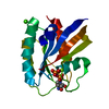
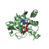
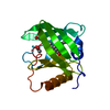
 Homo sapiens (human)
 X-RAY DIFFRACTION /
  Authors
 Citation
 Structure visualization
 Downloads & links
 Other downloads
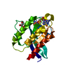


 PDBj
PDBj


 Assembly


 Mass: 24.305 Da / Num. of mol.: 1 / Source method: obtained synthetically / Formula: Mg
 Mass: 522.196 Da / Num. of mol.: 1 / Source method: obtained synthetically / Formula: C10H17N6O13P3
 Mass: 18.015 Da / Num. of mol.: 13 / Source method: isolated from a natural source / Formula: H2O
 Sample preparation
 Processing