 Movie
Movie Controller

[English] 日本語
 Yorodumi
- PDB-3ap6: Crystal structure of the galectin-8 N-terminal carbohydrate recog... -
+ Open data

- Basic information
| Entry | Database: PDB / ID: 3ap6 | |||||||||
|---|---|---|---|---|---|---|---|---|---|---|
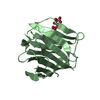
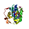
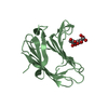
| Title | Crystal structure of the galectin-8 N-terminal carbohydrate recognition domain in complex with lactose 3'-sulfate | |||||||||
 Components Components | Galectin-8 | |||||||||
 Keywords Keywords | SUGAR BINDING PROTEIN / Beta-Sandwich / Galectin / Carbohydrate/Sugar Binding | |||||||||
| Function / homology |  Function and homology information Function and homology informationlymphatic endothelial cell migration / xenophagy / plasma cell differentiation / T cell costimulation / cellular response to virus / integrin binding / carbohydrate binding / cytoplasmic vesicle / : / membrane ...lymphatic endothelial cell migration / xenophagy / plasma cell differentiation / T cell costimulation / cellular response to virus / integrin binding / carbohydrate binding / cytoplasmic vesicle / : / membrane / cytoplasm / cytosol Similarity search - Function | |||||||||
| Biological species |  Homo sapiens (human) Homo sapiens (human) | |||||||||
| Method |  X-RAY DIFFRACTION / SYNCHROTRON / MOLECULAR REPLACEMENT / Resolution: 1.58 Å X-RAY DIFFRACTION / SYNCHROTRON / MOLECULAR REPLACEMENT / Resolution: 1.58 Å | |||||||||
 Authors Authors | Matsuzaka, T. / Ideo, H. / Yamashita, K. / Nonaka, T. | |||||||||
 Citation Citation | Journal: J.Biol.Chem. / Year: 2011 Title: Galectin-8-N-domain recognition mechanism for sialylated and sulfated glycans Authors: Ideo, H. / Matsuzaka, T. / Nonaka, T. / Seko, A. / Yamashita, K. | |||||||||
| History |
|
- Structure visualization
| Structure viewer | Molecule: MolmilJmol/JSmol |
|---|
- Downloads & links
-Download
| PDBx/mmCIF format | 3ap6.cif.gz | 141 KB | Display | PDBx/mmCIF format |
|---|---|---|---|---|
| PDB format | pdb3ap6.ent.gz | 109.3 KB | Display | PDB format |
| PDBx/mmJSON format | 3ap6.json.gz | Tree view | PDBx/mmJSON format | |
| Others |  Other downloads Other downloads |
-Validation report
| Arichive directory | https://data.pdbj.org/pub/pdb/validation_reports/ap/3ap6ftp://data.pdbj.org/pub/pdb/validation_reports/ap/3ap6 | HTTPS FTP |
|---|
-Related structure data
| Related structure data | 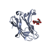 3ap4C  3ap5SC  3ap7C  3ap9C C: citing same article ( S: Starting model for refinement |
|---|---|
| Similar structure data |


-Links
 PDBj
PDBj


- Assembly
| Deposited unit | 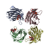
| ||||||||
|---|---|---|---|---|---|---|---|---|---|
| 1 | 
| ||||||||
| 2 | 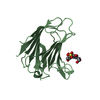
| ||||||||
| 3 | 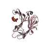
| ||||||||
| 4 | 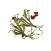
| ||||||||
| Unit cell |
|
-Components
| #1: Protein | Mass: 17423.123 Da / Num. of mol.: 4 / Fragment: N-terminal carbohydrate recognition domain Source method: isolated from a genetically manipulated source Source: (gene. exp.) Homo sapiens (human) / Gene: LGALS8 / Plasmid: pGEX-6P-2 / Production host:  #2: Polysaccharide | beta-D-galactopyranose-(1-4)-beta-D-glucopyranose / beta-lactose   Source method: isolated from a genetically manipulated source Details: oligosaccharide / References: beta-lactose #3: Chemical | ChemComp-SO4 /   Mass: 96.063 Da / Num. of mol.: 4 / Source method: obtained synthetically / Formula: SO4 Mass: 96.063 Da / Num. of mol.: 4 / Source method: obtained synthetically / Formula: SO4#4: Water | ChemComp-HOH / |  Mass: 18.015 Da / Num. of mol.: 491 / Source method: isolated from a natural source / Formula: H2O Mass: 18.015 Da / Num. of mol.: 491 / Source method: isolated from a natural source / Formula: H2OSequence details | THIS RESIDUE IS CAUSED BY NATURAL VARIANT. | |
|---|
-Experimental details
-Experiment
| Experiment | Method: X-RAY DIFFRACTION / Number of used crystals: 1 |
|---|
- Sample preparation
| Crystal | Density Matthews: 2.46 Å3/Da / Density % sol: 50.06 % |
|---|---|
| Crystal grow | Temperature: 293.15 K / Method: vapor diffusion, hanging drop / pH: 7 Details: 0.275mM protein, 10mM Hepes-NaOH, 2.75mM lactose 3'-sulfate, 50mM sodium chloride, 0.5mM DTT, 50mM ammonium fluoride, 9% PEG 3350, pH 7.0, VAPOR DIFFUSION, HANGING DROP, temperature 293.15K |
-Data collection
| Diffraction | Mean temperature: 95 K |
|---|---|
| Diffraction source | Source: SYNCHROTRON / Site: Photon Factory  / Beamline: BL-6A / Wavelength: 0.97798 Å / Beamline: BL-6A / Wavelength: 0.97798 Å |
| Detector | Type: ADSC QUANTUM 4r / Detector: CCD / Date: Oct 20, 2005 |
| Radiation | Monochromator: single crystal monochromator / Protocol: SINGLE WAVELENGTH / Monochromatic (M) / Laue (L): M / Scattering type: x-ray |
| Radiation wavelength | Wavelength: 0.97798 Å / Relative weight: 1 |
| Reflection | Resolution: 1.58→21.507 Å / Num. all: 86082 / Num. obs: 86082 / % possible obs: 94.4 % / Observed criterion σ(F): 0 / Observed criterion σ(I): 0 / Redundancy: 1.9 % / Biso Wilson estimate: 16.622 Å2 / Rmerge(I) obs: 0.055 / Rsym value: 0.055 |
| Reflection shell | Resolution: 1.58→1.62 Å / Redundancy: 1.9 % / Rmerge(I) obs: 0.121 / Mean I/σ(I) obs: 5.8 / Num. unique all: 6209 / Rsym value: 0.121 / % possible all: 91.5 |
- Processing
| Software |
| ||||||||||||||||||||||||||||||||||||||||||||||||||||||||||||||||||||||||||||||||||||||||||||||||||||||||||||||||||||||||||||||||||||||||||||||||||||||||||||||||||||||||||
|---|---|---|---|---|---|---|---|---|---|---|---|---|---|---|---|---|---|---|---|---|---|---|---|---|---|---|---|---|---|---|---|---|---|---|---|---|---|---|---|---|---|---|---|---|---|---|---|---|---|---|---|---|---|---|---|---|---|---|---|---|---|---|---|---|---|---|---|---|---|---|---|---|---|---|---|---|---|---|---|---|---|---|---|---|---|---|---|---|---|---|---|---|---|---|---|---|---|---|---|---|---|---|---|---|---|---|---|---|---|---|---|---|---|---|---|---|---|---|---|---|---|---|---|---|---|---|---|---|---|---|---|---|---|---|---|---|---|---|---|---|---|---|---|---|---|---|---|---|---|---|---|---|---|---|---|---|---|---|---|---|---|---|---|---|---|---|---|---|---|---|---|
| Refinement | Method to determine structure: MOLECULAR REPLACEMENT Starting model: PDB ENTRY 3AP5 Resolution: 1.58→21.44 Å / Cor.coef. Fo:Fc: 0.943 / Cor.coef. Fo:Fc free: 0.916 / SU B: 1.688 / SU ML: 0.062 / Isotropic thermal model: ISOTROPIC / Cross valid method: THROUGHOUT / ESU R: 0.132 / ESU R Free: 0.129 / Stereochemistry target values: Engh & Huber / Details: HYDROGENS HAVE BEEN ADDED IN THE RIDING POSITIONS
| ||||||||||||||||||||||||||||||||||||||||||||||||||||||||||||||||||||||||||||||||||||||||||||||||||||||||||||||||||||||||||||||||||||||||||||||||||||||||||||||||||||||||||
| Solvent computation | Ion probe radii: 0.8 Å / Shrinkage radii: 0.8 Å / VDW probe radii: 1.4 Å / Solvent model: BABINET MODEL WITH MASK | ||||||||||||||||||||||||||||||||||||||||||||||||||||||||||||||||||||||||||||||||||||||||||||||||||||||||||||||||||||||||||||||||||||||||||||||||||||||||||||||||||||||||||
| Displacement parameters | Biso mean: 15.907 Å2
| ||||||||||||||||||||||||||||||||||||||||||||||||||||||||||||||||||||||||||||||||||||||||||||||||||||||||||||||||||||||||||||||||||||||||||||||||||||||||||||||||||||||||||
| Refinement step | Cycle: LAST / Resolution: 1.58→21.44 Å
| ||||||||||||||||||||||||||||||||||||||||||||||||||||||||||||||||||||||||||||||||||||||||||||||||||||||||||||||||||||||||||||||||||||||||||||||||||||||||||||||||||||||||||
| Refine LS restraints |
| ||||||||||||||||||||||||||||||||||||||||||||||||||||||||||||||||||||||||||||||||||||||||||||||||||||||||||||||||||||||||||||||||||||||||||||||||||||||||||||||||||||||||||
| LS refinement shell | Resolution: 1.58→1.621 Å / Total num. of bins used: 20
|
