 Movie
Movie Controller

[English] 日本語
 Yorodumi
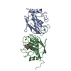
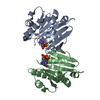
- PDB-1q3a: Crystal structure of the catalytic domain of human matrix metallo... -
+ Open data

- Basic information
| Entry | Database: PDB / ID: 1q3a | ||||||
|---|---|---|---|---|---|---|---|
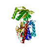
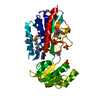
| Title | Crystal structure of the catalytic domain of human matrix metalloproteinase 10 | ||||||
 Components Components | Stromelysin-2 | ||||||
 Keywords Keywords | HYDROLASE / MMP-10 / metalloproteinase / inhibitors / NNGH / stromelysin-2 / hydroxamic acid | ||||||
| Function / homology |  Function and homology information Function and homology informationstromelysin 2 / Activation of Matrix Metalloproteinases / Collagen degradation / collagen catabolic process / extracellular matrix disassembly / Degradation of the extracellular matrix / extracellular matrix organization / metalloendopeptidase activity / extracellular matrix / serine-type endopeptidase activity ...stromelysin 2 / Activation of Matrix Metalloproteinases / Collagen degradation / collagen catabolic process / extracellular matrix disassembly / Degradation of the extracellular matrix / extracellular matrix organization / metalloendopeptidase activity / extracellular matrix / serine-type endopeptidase activity / proteolysis / : / extracellular region / zinc ion binding Similarity search - Function | ||||||
| Biological species |  Homo sapiens (human) Homo sapiens (human) | ||||||
| Method |  X-RAY DIFFRACTION / SYNCHROTRON / MOLECULAR REPLACEMENT / Resolution: 2.1 Å X-RAY DIFFRACTION / SYNCHROTRON / MOLECULAR REPLACEMENT / Resolution: 2.1 Å | ||||||
 Authors Authors | Calderone, V. / Bertini, I. / Fragai, M. / Luchinat, C. / Mangani, S. / Terni, B. | ||||||
 Citation Citation | Journal: J.Mol.Biol. / Year: 2004 Title: Crystal structure of the catalytic domain of human matrix metalloproteinase 10. Authors: Bertini, I. / Calderone, V. / Fragai, M. / Luchinat, C. / Mangani, S. / Terni, B. #1: Journal: J.Med.Chem. / Year: 2000Title: Development of New Hydroxamate Matrix Metalloproteinase Inhibitors Derived from Functionalized 4-Aminoprolines Authors: Natchus, M.G. / Bookland, R.G. / De, B. / Almstead, N.G. / Pikul, S. / Janusz, M.J. / Heitmeyer, S.A. / Hookfin, E.B. / Hsieh, L.C. / Dowty, M.E. / Dietsch, C.R. / Patel, V.S. / Garver, S.M. ...Authors: Natchus, M.G. / Bookland, R.G. / De, B. / Almstead, N.G. / Pikul, S. / Janusz, M.J. / Heitmeyer, S.A. / Hookfin, E.B. / Hsieh, L.C. / Dowty, M.E. / Dietsch, C.R. / Patel, V.S. / Garver, S.M. / Gu, F. / Pokross, M.E. / Mieling, G.E. / Baker, T.R. / Foltz, D.J. / Peng, S.X. / Bornes, D.M. / Strojnowski, M.J. / Taiwo, Y.O. #2: Journal: J.Med.Chem. / Year: 1999Title: Design, Synthesis, and Biological Evaluation of Potent Thiazine- and Thiazepine-Based Matrix Metalloproteinase Inhibitors Authors: Almstead, N.G. / Bradley, R.S. / Pikul, S. / De, B. / Natchus, M.G. / Taiwo, Y.O. / Gu, F. / Williams, L.E. / Hynd, B.A. / Janusz, M.J. / Dunaway, C.M. / Mieling, G.E. | ||||||
| History |
|
- Structure visualization
| Structure viewer | Molecule: MolmilJmol/JSmol |
|---|
- Downloads & links
-Download
| PDBx/mmCIF format | 1q3a.cif.gz | 121.6 KB | Display | PDBx/mmCIF format |
|---|---|---|---|---|
| PDB format | pdb1q3a.ent.gz | 93 KB | Display | PDB format |
| PDBx/mmJSON format | 1q3a.json.gz | Tree view | PDBx/mmJSON format | |
| Others |  Other downloads Other downloads |
-Validation report
| Arichive directory | https://data.pdbj.org/pub/pdb/validation_reports/q3/1q3aftp://data.pdbj.org/pub/pdb/validation_reports/q3/1q3a | HTTPS FTP |
|---|
-Related structure data
| Related structure data | 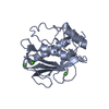 1g49S S: Starting model for refinement |
|---|---|
| Similar structure data |


-Links
 PDBj
PDBj


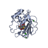
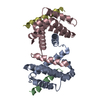
- Assembly
| Deposited unit | 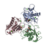
| ||||||||
|---|---|---|---|---|---|---|---|---|---|
| 1 | 
| ||||||||
| 2 | 
| ||||||||
| 3 | 
| ||||||||
| Unit cell |
| ||||||||
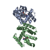
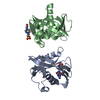
| Details | MMP-10 is physiologically a monomer but it forms a trimer in the crystallographic packing interactions in the asymmetric unit. |
-Components
| #1: Protein | Mass: 18537.488 Da / Num. of mol.: 3 / Fragment: catalytic domain / Mutation: F170N Source method: isolated from a genetically manipulated source Source: (gene. exp.) Homo sapiens (human) / Gene: MMP10 OR STMY2 / Production host:  #2: Chemical | ChemComp-ZN /   Mass: 65.409 Da / Num. of mol.: 6 / Source method: obtained synthetically / Formula: Zn Mass: 65.409 Da / Num. of mol.: 6 / Source method: obtained synthetically / Formula: Zn#3: Chemical | ChemComp-CA /   Mass: 40.078 Da / Num. of mol.: 9 / Source method: obtained synthetically / Formula: Ca Mass: 40.078 Da / Num. of mol.: 9 / Source method: obtained synthetically / Formula: Ca#4: Chemical |   Mass: 316.373 Da / Num. of mol.: 3 / Source method: obtained synthetically / Formula: C13H20N2O5S Mass: 316.373 Da / Num. of mol.: 3 / Source method: obtained synthetically / Formula: C13H20N2O5S#5: Water | ChemComp-HOH / |  Mass: 18.015 Da / Num. of mol.: 351 / Source method: isolated from a natural source / Formula: H2O Mass: 18.015 Da / Num. of mol.: 351 / Source method: isolated from a natural source / Formula: H2O |
|---|
-Experimental details
-Experiment
| Experiment | Method: X-RAY DIFFRACTION / Number of used crystals: 1 |
|---|
- Sample preparation
| Crystal | Density Matthews: 2.09 Å3/Da / Density % sol: 41.18 % |
|---|---|
| Crystal grow | Temperature: 293 K / Method: vapor diffusion, sitting drop / pH: 8 Details: Tris-HCl, PEG 6000, Acetohydroxamic acid, NNGH, pH 8.0, VAPOR DIFFUSION, SITTING DROP, temperature 293K |
-Data collection
| Diffraction | Mean temperature: 100 K |
|---|---|
| Diffraction source | Source: SYNCHROTRON / Site: ESRF  / Beamline: ID29 / Wavelength: 0.933 Å / Beamline: ID29 / Wavelength: 0.933 Å |
| Detector | Type: ADSC QUANTUM 4 / Detector: CCD / Date: Apr 4, 2003 / Details: Si(111) |
| Radiation | Monochromator: Si(111) / Protocol: SINGLE WAVELENGTH / Monochromatic (M) / Laue (L): M / Scattering type: x-ray |
| Radiation wavelength | Wavelength: 0.933 Å / Relative weight: 1 |
| Reflection | Resolution: 2.1→20 Å / Num. obs: 25246 / Observed criterion σ(F): 0 / Observed criterion σ(I): 0 / Redundancy: 3 % / Rmerge(I) obs: 0.14 / Rsym value: 0.14 / Net I/σ(I): 3.6 |
| Reflection shell | Resolution: 2.1→2.21 Å / Redundancy: 2.1 % / Rmerge(I) obs: 0.38 / Mean I/σ(I) obs: 1.8 / Rsym value: 0.38 / % possible all: 89.1 |
- Processing
| Software |
| ||||||||||||||||||||||||||||||||||||||||||||||||||||||||||||||||||||||||||||||||||||||||||||||||||||||||||||||||||||||||||||||||||||||||||||||||||||||||||||||||
|---|---|---|---|---|---|---|---|---|---|---|---|---|---|---|---|---|---|---|---|---|---|---|---|---|---|---|---|---|---|---|---|---|---|---|---|---|---|---|---|---|---|---|---|---|---|---|---|---|---|---|---|---|---|---|---|---|---|---|---|---|---|---|---|---|---|---|---|---|---|---|---|---|---|---|---|---|---|---|---|---|---|---|---|---|---|---|---|---|---|---|---|---|---|---|---|---|---|---|---|---|---|---|---|---|---|---|---|---|---|---|---|---|---|---|---|---|---|---|---|---|---|---|---|---|---|---|---|---|---|---|---|---|---|---|---|---|---|---|---|---|---|---|---|---|---|---|---|---|---|---|---|---|---|---|---|---|---|---|---|---|---|
| Refinement | Method to determine structure: MOLECULAR REPLACEMENT Starting model: PDB entry 1G49 Resolution: 2.1→51.3 Å / SU B: 9.54 / SU ML: 0.238 / Isotropic thermal model: RESTRAINED / Cross valid method: THROUGHOUT / ESU R: 0.539 / ESU R Free: 0.282 / Stereochemistry target values: MAXIMUM LIKELIHOOD
| ||||||||||||||||||||||||||||||||||||||||||||||||||||||||||||||||||||||||||||||||||||||||||||||||||||||||||||||||||||||||||||||||||||||||||||||||||||||||||||||||
| Solvent computation | Ion probe radii: 0.8 Å / Shrinkage radii: 0.8 Å / VDW probe radii: 1.4 Å / Solvent model: BABINET MODEL WITH MASK | ||||||||||||||||||||||||||||||||||||||||||||||||||||||||||||||||||||||||||||||||||||||||||||||||||||||||||||||||||||||||||||||||||||||||||||||||||||||||||||||||
| Displacement parameters | Biso mean: 37.771 Å2
| ||||||||||||||||||||||||||||||||||||||||||||||||||||||||||||||||||||||||||||||||||||||||||||||||||||||||||||||||||||||||||||||||||||||||||||||||||||||||||||||||
| Refine analyze | Luzzati coordinate error free: 0.33 Å / Luzzati sigma a free: 0.33 Å | ||||||||||||||||||||||||||||||||||||||||||||||||||||||||||||||||||||||||||||||||||||||||||||||||||||||||||||||||||||||||||||||||||||||||||||||||||||||||||||||||
| Refinement step | Cycle: LAST / Resolution: 2.1→51.3 Å
| ||||||||||||||||||||||||||||||||||||||||||||||||||||||||||||||||||||||||||||||||||||||||||||||||||||||||||||||||||||||||||||||||||||||||||||||||||||||||||||||||
| Refine LS restraints |
| ||||||||||||||||||||||||||||||||||||||||||||||||||||||||||||||||||||||||||||||||||||||||||||||||||||||||||||||||||||||||||||||||||||||||||||||||||||||||||||||||
| LS refinement shell | Resolution: 2.1→2.155 Å / Total num. of bins used: 20 /
| ||||||||||||||||||||||||||||||||||||||||||||||||||||||||||||||||||||||||||||||||||||||||||||||||||||||||||||||||||||||||||||||||||||||||||||||||||||||||||||||||
| Xplor file |
|
