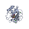
 Movie
Movie Controller


+ Open data

- Basic information
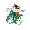
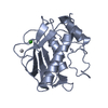
| Entry | Database: PDB / ID: 1g49 | ||||||
|---|---|---|---|---|---|---|---|
| Title | A CARBOXYLIC ACID BASED INHIBITOR IN COMPLEX WITH MMP3 | ||||||
 Components Components | MATRIX METALLOPROTEINASE 3 | ||||||
 Keywords Keywords | HYDROLASE / mixed alpha beta structure / Zinc protease / inhibited | ||||||
| Function / homology |  Function and homology information Function and homology informationstromelysin 1 / cellular response to UV-A / regulation of neuroinflammatory response / Assembly of collagen fibrils and other multimeric structures / Activation of Matrix Metalloproteinases / response to amyloid-beta / Collagen degradation / collagen catabolic process / negative regulation of reactive oxygen species metabolic process / extracellular matrix disassembly ...stromelysin 1 / cellular response to UV-A / regulation of neuroinflammatory response / Assembly of collagen fibrils and other multimeric structures / Activation of Matrix Metalloproteinases / response to amyloid-beta / Collagen degradation / collagen catabolic process / negative regulation of reactive oxygen species metabolic process / extracellular matrix disassembly / Degradation of the extracellular matrix / extracellular matrix organization / cellular response to nitric oxide / EGFR Transactivation by Gastrin / regulation of cell migration / negative regulation of phosphatidylinositol 3-kinase/protein kinase B signal transduction / protein catabolic process / cellular response to reactive oxygen species / cellular response to amino acid stimulus / positive regulation of protein-containing complex assembly / metalloendopeptidase activity / metallopeptidase activity / peptidase activity / extracellular matrix / cellular response to lipopolysaccharide / Interleukin-4 and Interleukin-13 signaling / endopeptidase activity / Extra-nuclear estrogen signaling / serine-type endopeptidase activity / innate immune response / mitochondrion / proteolysis / : / extracellular region / zinc ion binding / nucleus / cytosol Similarity search - Function | ||||||
| Biological species |  Homo sapiens (human) Homo sapiens (human) | ||||||
| Method |  X-RAY DIFFRACTION / SYNCHROTRON / FOURIER SYNTHESIS / Resolution: 1.9 Å X-RAY DIFFRACTION / SYNCHROTRON / FOURIER SYNTHESIS / Resolution: 1.9 Å | ||||||
 Authors Authors | Natchus, M.G. / Bookland, R.G. / De, B. / Almstead, N.G. / Pikul, S. | ||||||
 Citation Citation | Journal: J.Med.Chem. / Year: 2000 Title: Development of new hydroxamate matrix metalloproteinase inhibitors derived from functionalized 4-aminoprolines. Authors: Natchus, M.G. / Bookland, R.G. / De, B. / Almstead, N.G. / Pikul, S. | ||||||
| History |
|
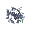
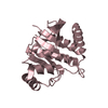
- Structure visualization
| Structure viewer | Molecule: MolmilJmol/JSmol |
|---|
- Downloads & links
-Download
| PDBx/mmCIF format | 1g49.cif.gz | 86.3 KB | Display | PDBx/mmCIF format |
|---|---|---|---|---|
| PDB format | pdb1g49.ent.gz | 64.2 KB | Display | PDB format |
| PDBx/mmJSON format | 1g49.json.gz | Tree view | PDBx/mmJSON format | |
| Others |  Other downloads Other downloads |
-Validation report
| Arichive directory | https://data.pdbj.org/pub/pdb/validation_reports/g4/1g49ftp://data.pdbj.org/pub/pdb/validation_reports/g4/1g49 | HTTPS FTP |
|---|
-Related structure data
| Related structure data | |
|---|---|
| Similar structure data |


-Links
 PDBj
PDBj


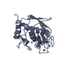
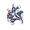
- Assembly
| Deposited unit | 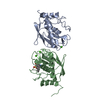
| ||||||||||
|---|---|---|---|---|---|---|---|---|---|---|---|
| 1 | 
| ||||||||||
| 2 | 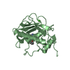
| ||||||||||
| Unit cell |
| ||||||||||
| Details | The biological assembly is a monomer constructed from chain A or chain B |
-Components
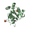
| #1: Protein | Mass: 19416.529 Da / Num. of mol.: 2 / Fragment: CATALYTIC DOMAIN Source method: isolated from a genetically manipulated source Source: (gene. exp.) Homo sapiens (human) / Cell: FIBROBLAST / Gene: MMP3 / Production host:  #2: Chemical | ChemComp-ZN /   Mass: 65.409 Da / Num. of mol.: 4 / Source method: obtained synthetically / Formula: Zn Mass: 65.409 Da / Num. of mol.: 4 / Source method: obtained synthetically / Formula: Zn#3: Chemical | ChemComp-CA /   Mass: 40.078 Da / Num. of mol.: 6 / Source method: obtained synthetically / Formula: Ca Mass: 40.078 Da / Num. of mol.: 6 / Source method: obtained synthetically / Formula: Ca#4: Chemical | ChemComp-111 / ( |   Mass: 435.516 Da / Num. of mol.: 1 / Source method: obtained synthetically / Formula: C16H25N3O7S2 Mass: 435.516 Da / Num. of mol.: 1 / Source method: obtained synthetically / Formula: C16H25N3O7S2#5: Water | ChemComp-HOH / |  Mass: 18.015 Da / Num. of mol.: 111 / Source method: isolated from a natural source / Formula: H2O Mass: 18.015 Da / Num. of mol.: 111 / Source method: isolated from a natural source / Formula: H2O |
|---|
-Experimental details
-Experiment
| Experiment | Method: X-RAY DIFFRACTION / Number of used crystals: 1 |
|---|
- Sample preparation
| Crystal | Density Matthews: 1.98 Å3/Da / Density % sol: 37.99 % | ||||||||||||||||||||||||||||||||||||
|---|---|---|---|---|---|---|---|---|---|---|---|---|---|---|---|---|---|---|---|---|---|---|---|---|---|---|---|---|---|---|---|---|---|---|---|---|---|
| Crystal grow | Temperature: 298 K / Method: vapor diffusion, hanging drop / pH: 8.5 Details: PEG 6000, NaCl, MgCl2, Tris, pH 8.5, VAPOR DIFFUSION, HANGING DROP at 298K | ||||||||||||||||||||||||||||||||||||
| Crystal grow | *PLUS Method: vapor diffusionDetails: used macroseeding, Chen, L., (1999) J. Mol. Biol., 293, 545. PH range low: 8.5 / PH range high: 7.5 | ||||||||||||||||||||||||||||||||||||
| Components of the solutions | *PLUS
|
-Data collection
| Diffraction | Mean temperature: 173 K |
|---|---|
| Diffraction source | Source: SYNCHROTRON / Site: APS  / Beamline: 17-ID / Wavelength: 1 Å / Beamline: 17-ID / Wavelength: 1 Å |
| Detector | Type: MARRESEARCH / Detector: CCD / Date: Mar 30, 2000 / Details: monochromator |
| Radiation | Protocol: SINGLE WAVELENGTH / Monochromatic (M) / Laue (L): M / Scattering type: x-ray |
| Radiation wavelength | Wavelength: 1 Å / Relative weight: 1 |
| Reflection | Resolution: 1.8→62.1 Å / Num. all: 30250 / Num. obs: 22034 / % possible obs: 72.8 % / Redundancy: 1.6 % / Biso Wilson estimate: 15.2 Å2 / Rmerge(I) obs: 0.065 / Net I/σ(I): 7.4 |
| Reflection shell | Resolution: 1.8→1.85 Å / Redundancy: 1.3 % / Rmerge(I) obs: 0.225 / Num. unique all: 1726 / % possible all: 30.7 |
- Processing
| Software |
| ||||||||||||||||||||
|---|---|---|---|---|---|---|---|---|---|---|---|---|---|---|---|---|---|---|---|---|---|
| Refinement | Method to determine structure: FOURIER SYNTHESIS / Resolution: 1.9→19.7 Å / Isotropic thermal model: anisotropic / Cross valid method: Free R / Stereochemistry target values: Engh & Huber / Details: Used maximum likelihood procedures
| ||||||||||||||||||||
| Displacement parameters | Biso mean: 24 Å2
| ||||||||||||||||||||
| Refinement step | Cycle: LAST / Resolution: 1.9→19.7 Å
| ||||||||||||||||||||
| Refine LS restraints |
| ||||||||||||||||||||
| Software | *PLUS Name: REFMAC / Classification: refinement | ||||||||||||||||||||
| Refinement | *PLUS Highest resolution: 1.9 Å / % reflection Rfree: 11 % / Rfactor Rwork: 0.196 | ||||||||||||||||||||
| Solvent computation | *PLUS | ||||||||||||||||||||
| Displacement parameters | *PLUS Biso mean: 24 Å2 | ||||||||||||||||||||
| Refine LS restraints | *PLUS Type: p_angle_d / Dev ideal: 1.9 |
