| 登録情報 | データベース: PDB / ID: 1cqr
|
|---|
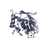
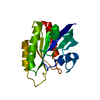
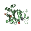
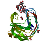
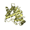
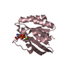
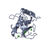
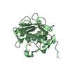
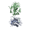
| タイトル | CRYSTAL STRUCTURE OF THE STROMELYSIN CATALYTIC DOMAIN AT 2.0 A RESOLUTION |
|---|
 要素 要素 | STROMELYSIN-1 |
|---|
 キーワード キーワード | HYDROLASE / ACTIVE / TRUNCATED NATIVE ENZYME |
|---|
| 機能・相同性 |  機能・相同性情報 機能・相同性情報
stromelysin 1 / cellular response to UV-A / regulation of neuroinflammatory response / Assembly of collagen fibrils and other multimeric structures / Activation of Matrix Metalloproteinases / response to amyloid-beta / Collagen degradation / collagen catabolic process / negative regulation of reactive oxygen species metabolic process / extracellular matrix disassembly ...stromelysin 1 / cellular response to UV-A / regulation of neuroinflammatory response / Assembly of collagen fibrils and other multimeric structures / Activation of Matrix Metalloproteinases / response to amyloid-beta / Collagen degradation / collagen catabolic process / negative regulation of reactive oxygen species metabolic process / extracellular matrix disassembly / Degradation of the extracellular matrix / extracellular matrix organization / cellular response to nitric oxide / EGFR Transactivation by Gastrin / regulation of cell migration / protein catabolic process / negative regulation of phosphatidylinositol 3-kinase/protein kinase B signal transduction / cellular response to amino acid stimulus / cellular response to reactive oxygen species / positive regulation of protein-containing complex assembly / metalloendopeptidase activity / metallopeptidase activity / peptidase activity / cellular response to lipopolysaccharide / extracellular matrix / Interleukin-4 and Interleukin-13 signaling / endopeptidase activity / Extra-nuclear estrogen signaling / serine-type endopeptidase activity / innate immune response / mitochondrion / proteolysis / : / extracellular region / zinc ion binding / nucleus / cytosol類似検索 - 分子機能 Hemopexin, conserved site / Hemopexin domain signature. / Hemopexin-like domain / Peptidase M10A, cysteine switch, zinc binding site / Matrixins cysteine switch. / Peptidoglycan binding-like / Hemopexin-like repeats / Hemopexin-like domain superfamily / Hemopexin / Hemopexin repeat profile. ...Hemopexin, conserved site / Hemopexin domain signature. / Hemopexin-like domain / Peptidase M10A, cysteine switch, zinc binding site / Matrixins cysteine switch. / Peptidoglycan binding-like / Hemopexin-like repeats / Hemopexin-like domain superfamily / Hemopexin / Hemopexin repeat profile. / Hemopexin-like repeats. / Peptidase M10A / Peptidase M10A, catalytic domain / Putative peptidoglycan binding domain / Peptidase M10, metallopeptidase / Matrixin / PGBD-like superfamily / Peptidase, metallopeptidase / Zinc-dependent metalloprotease / Collagenase (Catalytic Domain) / Collagenase (Catalytic Domain) / Metallopeptidase, catalytic domain superfamily / Neutral zinc metallopeptidases, zinc-binding region signature. / 3-Layer(aba) Sandwich / Alpha Beta類似検索 - ドメイン・相同性 |
|---|
| 生物種 |  Homo sapiens (ヒト) Homo sapiens (ヒト) |
|---|
| 手法 |  X線回折 / 解像度: 2 Å X線回折 / 解像度: 2 Å |
|---|
 データ登録者 データ登録者 | Chen, L. / Rydel, T.J. / Gu, F. / Dunaway, C.M. / Pikul, S. / Dunham, K.M. / Barnett, B.L. |
|---|
 引用 引用 | #1: ジャーナル: J.Med.Chem. / 年: 1999タイトル: Design and Synthesis of Phosphinamide-Based Hydroxamic Acids as Inhibitors of Matrix Metalloproteinases 著者: Pikul, S. / McDow Dunham, K.L. / Almstead, N.G. / De, B. / Natchus, M.G. / Anastasio, M.V. / McPhail, S.J. / Snider, C.E. / Taiwo, Y.O. / Chen, L. / Dunaway, C.M. / Gu, F. / Mieling, G.E. |
|---|
| 履歴 | | 登録 | 1999年8月11日 | 登録サイト: RCSB / 処理サイト: RCSB |
|---|
| 改定 1.0 | 2000年3月20日 | Provider: repository / タイプ: Initial release |
|---|
| 改定 1.1 | 2008年4月27日 | Group: Version format compliance |
|---|
| 改定 1.2 | 2011年7月13日 | Group: Derived calculations / Version format compliance |
|---|
| 改定 1.3 | 2024年2月7日 | Group: Data collection / Database references / Derived calculations
カテゴリ: chem_comp_atom / chem_comp_bond ...chem_comp_atom / chem_comp_bond / database_2 / pdbx_struct_conn_angle / struct_conn / struct_site
Item: _database_2.pdbx_DOI / _database_2.pdbx_database_accession ..._database_2.pdbx_DOI / _database_2.pdbx_database_accession / _pdbx_struct_conn_angle.ptnr1_auth_asym_id / _pdbx_struct_conn_angle.ptnr1_auth_comp_id / _pdbx_struct_conn_angle.ptnr1_auth_seq_id / _pdbx_struct_conn_angle.ptnr1_label_asym_id / _pdbx_struct_conn_angle.ptnr1_label_atom_id / _pdbx_struct_conn_angle.ptnr1_label_comp_id / _pdbx_struct_conn_angle.ptnr1_label_seq_id / _pdbx_struct_conn_angle.ptnr1_symmetry / _pdbx_struct_conn_angle.ptnr2_auth_comp_id / _pdbx_struct_conn_angle.ptnr2_auth_seq_id / _pdbx_struct_conn_angle.ptnr2_label_asym_id / _pdbx_struct_conn_angle.ptnr2_label_atom_id / _pdbx_struct_conn_angle.ptnr2_label_comp_id / _pdbx_struct_conn_angle.ptnr3_auth_asym_id / _pdbx_struct_conn_angle.ptnr3_auth_comp_id / _pdbx_struct_conn_angle.ptnr3_auth_seq_id / _pdbx_struct_conn_angle.ptnr3_label_asym_id / _pdbx_struct_conn_angle.ptnr3_label_atom_id / _pdbx_struct_conn_angle.ptnr3_label_comp_id / _pdbx_struct_conn_angle.ptnr3_label_seq_id / _pdbx_struct_conn_angle.ptnr3_symmetry / _pdbx_struct_conn_angle.value / _struct_conn.pdbx_dist_value / _struct_conn.ptnr1_auth_asym_id / _struct_conn.ptnr1_auth_comp_id / _struct_conn.ptnr1_auth_seq_id / _struct_conn.ptnr1_label_asym_id / _struct_conn.ptnr1_label_atom_id / _struct_conn.ptnr1_label_comp_id / _struct_conn.ptnr1_label_seq_id / _struct_conn.ptnr2_auth_comp_id / _struct_conn.ptnr2_auth_seq_id / _struct_conn.ptnr2_label_asym_id / _struct_conn.ptnr2_label_atom_id / _struct_conn.ptnr2_label_comp_id / _struct_conn.ptnr2_label_seq_id / _struct_conn.ptnr2_symmetry / _struct_site.pdbx_auth_asym_id / _struct_site.pdbx_auth_comp_id / _struct_site.pdbx_auth_seq_id |
|---|
|
|---|
 ムービー
ムービー コントローラー


 データを開く
 基本情報
基本情報 構造の表示
構造の表示 ダウンロードとリンク
 その他のダウンロード


 PDBj
PDBj


 集合体
集合体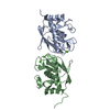


 分子量: 65.409 Da / 分子数: 4 / 由来タイプ: 合成 / 式: Zn
 分子量: 40.078 Da / 分子数: 6 / 由来タイプ: 合成 / 式: Ca
 分子量: 18.015 Da / 分子数: 82 / 由来タイプ: 天然 / 式: H2O
 試料調製
試料調製 解析
解析