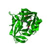
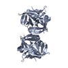
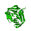
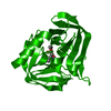
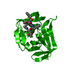
[English] 日本語
Yorodumi
- PDB-3zz6: Crystal structure of 3C protease of coxsackievirus B3 complexed w... -

+
Open data


ID or keywords:

Loading...

-
Basic information

Entry
Database: PDB / ID: 3zz6
TitleCrystal structure of 3C protease of coxsackievirus B3 complexed with Michael receptor inhibitor 75
ComponentsPOLYPROTEIN 3BCD
KeywordsHYDROLASE / PICORNAVIRIDAE / MICHAEL RECEPTOR INHIBITOR
Function / homology
Function and homology information


symbiont-mediated suppression of host cytoplasmic pattern recognition receptor signaling pathway via inhibition of RIG-I activity / picornain 2A / symbiont-mediated suppression of host mRNA export from nucleus / symbiont genome entry into host cell via pore formation in plasma membrane / picornain 3C / T=pseudo3 icosahedral viral capsid / host cell cytoplasmic vesicle membrane / endocytosis involved in viral entry into host cell / cytoplasmic vesicle membrane / : ...symbiont-mediated suppression of host cytoplasmic pattern recognition receptor signaling pathway via inhibition of RIG-I activity / picornain 2A / symbiont-mediated suppression of host mRNA export from nucleus / symbiont genome entry into host cell via pore formation in plasma membrane / picornain 3C / T=pseudo3 icosahedral viral capsid / host cell cytoplasmic vesicle membrane / endocytosis involved in viral entry into host cell / cytoplasmic vesicle membrane / : / nucleoside-triphosphate phosphatase / protein complex oligomerization / monoatomic ion channel activity / host cell cytoplasm / RNA helicase activity / DNA replication / induction by virus of host autophagy / RNA-directed RNA polymerase / symbiont entry into host cell / symbiont-mediated suppression of host gene expression / viral RNA genome replication / cysteine-type endopeptidase activity / RNA-dependent RNA polymerase activity / nucleotide binding / DNA-templated transcription / host cell nucleus / structural molecule activity / virion attachment to host cell / ATP hydrolysis activity / proteolysis / RNA binding / ATP binding / metal ion binding / cytoplasm
Similarity search - Function
Picornavirus coat protein VP4 superfamily / Poliovirus 3A protein-like / Poliovirus 3A protein like / Picornavirus 2B protein / Poliovirus core protein 3a, soluble domain / Picornavirus 2B protein / Peptidase C3, picornavirus core protein 2A / Picornavirus core protein 2A / Picornavirus coat protein VP4 / Picornavirus coat protein (VP4) ...Picornavirus coat protein VP4 superfamily / Poliovirus 3A protein-like / Poliovirus 3A protein like / Picornavirus 2B protein / Poliovirus core protein 3a, soluble domain / Picornavirus 2B protein / Peptidase C3, picornavirus core protein 2A / Picornavirus core protein 2A / Picornavirus coat protein VP4 / Picornavirus coat protein (VP4) / Picornavirales 3C/3C-like protease domain / Picornavirales 3C/3C-like protease domain profile. / Peptidase C3A/C3B, picornaviral / 3C cysteine protease (picornain 3C) / Picornavirus capsid / picornavirus capsid protein / Helicase, superfamily 3, single-stranded RNA virus / Superfamily 3 helicase of positive ssRNA viruses domain profile. / Helicase, superfamily 3, single-stranded DNA/RNA virus / RNA helicase / Picornavirus/Calicivirus coat protein / Viral coat protein subunit / Trypsin-like serine proteases / Thrombin, subunit H / RNA-directed RNA polymerase, C-terminal domain / Viral RNA-dependent RNA polymerase / Reverse transcriptase/Diguanylate cyclase domain / RNA-directed RNA polymerase, catalytic domain / RdRp of positive ssRNA viruses catalytic domain profile. / ATPases associated with a variety of cellular activities / AAA+ ATPase domain / Peptidase S1, PA clan, chymotrypsin-like fold / DNA/RNA polymerase superfamily / Peptidase S1, PA clan / Beta Barrel / P-loop containing nucleoside triphosphate hydrolase / Mainly Beta
Similarity search - Domain/homology
Chem-G75 / Genome polyprotein / Genome polyprotein
Similarity search - Component
Biological speciesHUMAN COXSACKIEVIRUS B3
MethodX-RAY DIFFRACTION / SYNCHROTRON / MOLECULAR REPLACEMENT / Resolution: 2.05 Å
AuthorsTan, J. / Anand, K. / Mesters, J.R. / Hilgenfeld, R.
CitationJournal: To be Published
Title: Peptidic Ab-Nonsaturated Ethyl Esters as Inhibitors of the 3C Protease of Coxsackie Virus B3: Crystal Structures, Antiviral Activities, and Resistance Mutations
Authors: Tan, J. / Anand, K. / Mesters, J.R. / Hilgenfeld, R.
History
DepositionAug 31, 2011Deposition site: PDBE / Processing site: PDBE
Revision 1.0Sep 19, 2012Provider: repository / Type: Initial release
Revision 1.1May 8, 2019Group: Data collection / Derived calculations ...Data collection / Derived calculations / Experimental preparation / Other
Category: exptl_crystal_grow / pdbx_database_proc ...exptl_crystal_grow / pdbx_database_proc / pdbx_database_status / struct_conn
Item: _exptl_crystal_grow.method / _pdbx_database_status.recvd_author_approval / _struct_conn.pdbx_leaving_atom_flag
Remark 700 SHEET DETERMINATION METHOD: DSSP THE SHEETS PRESENTED AS "AA" IN EACH CHAIN ON SHEET RECORDS BELOW ... SHEET DETERMINATION METHOD: DSSP THE SHEETS PRESENTED AS "AA" IN EACH CHAIN ON SHEET RECORDS BELOW IS ACTUALLY AN 6-STRANDED BARREL THIS IS REPRESENTED BY A 7-STRANDED SHEET IN WHICH THE FIRST AND LAST STRANDS ARE IDENTICAL. THE SHEETS PRESENTED AS "AB" IN EACH CHAIN ON SHEET RECORDS BELOW IS ACTUALLY AN 6-STRANDED BARREL THIS IS REPRESENTED BY A 7-STRANDED SHEET IN WHICH THE FIRST AND LAST STRANDS ARE IDENTICAL.

-
Structure visualization

Structure viewerMolecule:
MolmilJmol/JSmol

Downloads & links

-
Assembly

Deposited unit
A: POLYPROTEIN 3BCD
hetero molecules


Theoretical massNumber of molelcules
Total (without water)20,9392
Polymers20,4291
Non-polymers5101
Water61334
1
A: POLYPROTEIN 3BCD
hetero molecules

A: POLYPROTEIN 3BCD
hetero molecules


Theoretical massNumber of molelcules
Total (without water)41,8784
Polymers40,8592
Non-polymers1,0192
Water362
TypeNameSymmetry operationNumber
identity operation1_555x,y,z1
crystal symmetry operation2_454-x-1,y,-z-11
Buried area1740 Å2
ΔGint-15.4 kcal/mol
Surface area14970 Å2
MethodPISA
Unit cell
Length a, b, c (Å)76.920, 64.090, 39.540
Angle α, β, γ (deg.)90.00, 116.63, 90.00
Int Tables number5
Space group name H-MC121

-
Components

#1: Protein POLYPROTEIN 3BCD / 3C PROTEASE OF COXSACKIEVIRUS B3


Mass: 20429.488 Da / Num. of mol.: 1
Source method: isolated from a genetically manipulated source
Source: (gene. exp.) HUMAN COXSACKIEVIRUS B3 / Strain: COXSACKIEVIRUS B3 / Plasmid: PET23A-COX / Production host: ESCHERICHIA COLI (E. coli) / Strain (production host): BL21(DE3)GOLD
References: UniProt: Q90092, UniProt: Q5UEA2*PLUS, picornain 3C
#2: Chemical ChemComp-G75 / ETHYL (4R)-4-({N-[(BENZYLOXY)CARBONYL]-L-PHENYLALANYL}AMINO)-5-[(3S)-2-OXOPYRROLIDIN-3-YL]PENTANOATE


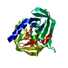
Mass: 509.594 Da / Num. of mol.: 1 / Source method: obtained synthetically / Formula: C28H35N3O6
#3: Water ChemComp-HOH / water / Water


Mass: 18.015 Da / Num. of mol.: 34 / Source method: isolated from a natural source / Formula: H2O

-
Experimental details

-
Experiment

ExperimentMethod: X-RAY DIFFRACTION / Number of used crystals: 1

-
Sample preparation

CrystalDensity Matthews: 2.07 Å3/Da / Density % sol: 40.74 % / Description: NONE
Crystal growMethod: vapor diffusion, sitting drop / pH: 8.5
Details: 100 MM TRIS-HCL PH 8.5, 0.2 M MAGNESIUM CHLORIDE, AND 22% PEG 4000; SITTING DROP

-
Data collection

DiffractionMean temperature: 100 K
Diffraction sourceSource: SYNCHROTRON / Site: BESSY / Beamline: 14.1 / Wavelength: 0.9184
DetectorType: MARRESEARCH / Detector: CCD / Details: MIRRORS
RadiationProtocol: SINGLE WAVELENGTH / Monochromatic (M) / Laue (L): M / Scattering type: x-ray
Radiation wavelengthWavelength: 0.9184 Å / Relative weight: 1
ReflectionResolution: 2.05→35.35 Å / Num. obs: 10746 / % possible obs: 99.3 % / Observed criterion σ(I): 2 / Redundancy: 3.6 % / Rmerge(I) obs: 0.06 / Net I/σ(I): 9.3
Reflection shellResolution: 2.05→2.16 Å / Redundancy: 3.6 % / Rmerge(I) obs: 0.44 / Mean I/σ(I) obs: 2.4 / % possible all: 98.9

-
Processing

Software
NameVersionClassification
REFMAC5.5.0110refinement
iMOSFLMdata reduction
SCALAdata scaling
MOLREPphasing
RefinementMethod to determine structure: MOLECULAR REPLACEMENT
Starting model: CRYSTAL STRUCTURE OF COXSACKIEVIURS B3 3C PROTEASE

Resolution: 2.05→46.88 Å / Cor.coef. Fo:Fc: 0.952 / Cor.coef. Fo:Fc free: 0.914 / SU B: 7.728 / SU ML: 0.21 / Cross valid method: THROUGHOUT / ESU R: 0.275 / ESU R Free: 0.226 / Stereochemistry target values: MAXIMUM LIKELIHOOD
Details: HYDROGENS HAVE BEEN ADDED IN THE RIDING POSITIONS.U VALUES REFINED INDIVIDUALLY
RfactorNum. reflection% reflectionSelection details
Rfree0.28484 516 4.8 %RANDOM
Rwork0.22517 ---
obs0.22806 10230 99.04 %-
Solvent computationIon probe radii: 0.8 Å / Shrinkage radii: 0.8 Å / VDW probe radii: 1.4 Å / Solvent model: MASK
Displacement parametersBiso mean: 45.471 Å2
Baniso -1Baniso -2Baniso -3
1--0.1 Å20 Å22.64 Å2
2---0.82 Å20 Å2
3---3.29 Å2
Refinement stepCycle: LAST / Resolution: 2.05→46.88 Å
ProteinNucleic acidLigandSolventTotal
Num. atoms1406 0 37 34 1477
Refine LS restraints
Refine-IDTypeDev idealDev ideal targetNumber
X-RAY DIFFRACTIONr_bond_refined_d0.0180.0221475
X-RAY DIFFRACTIONr_bond_other_d
X-RAY DIFFRACTIONr_angle_refined_deg1.8861.9931985
X-RAY DIFFRACTIONr_angle_other_deg
X-RAY DIFFRACTIONr_dihedral_angle_1_deg7.3385180
X-RAY DIFFRACTIONr_dihedral_angle_2_deg32.32724.12763
X-RAY DIFFRACTIONr_dihedral_angle_3_deg18.63915252
X-RAY DIFFRACTIONr_dihedral_angle_4_deg13.363158
X-RAY DIFFRACTIONr_chiral_restr0.110.2215
X-RAY DIFFRACTIONr_gen_planes_refined0.0080.0211112
X-RAY DIFFRACTIONr_gen_planes_other
X-RAY DIFFRACTIONr_nbd_refined
X-RAY DIFFRACTIONr_nbd_other
X-RAY DIFFRACTIONr_nbtor_refined
X-RAY DIFFRACTIONr_nbtor_other
X-RAY DIFFRACTIONr_xyhbond_nbd_refined
X-RAY DIFFRACTIONr_xyhbond_nbd_other
X-RAY DIFFRACTIONr_metal_ion_refined
X-RAY DIFFRACTIONr_metal_ion_other
X-RAY DIFFRACTIONr_symmetry_vdw_refined
X-RAY DIFFRACTIONr_symmetry_vdw_other
X-RAY DIFFRACTIONr_symmetry_hbond_refined
X-RAY DIFFRACTIONr_symmetry_hbond_other
X-RAY DIFFRACTIONr_symmetry_metal_ion_refined
X-RAY DIFFRACTIONr_symmetry_metal_ion_other
X-RAY DIFFRACTIONr_mcbond_it1.1431.5892
X-RAY DIFFRACTIONr_mcbond_other
X-RAY DIFFRACTIONr_mcangle_it2.01321429
X-RAY DIFFRACTIONr_mcangle_other
X-RAY DIFFRACTIONr_scbond_it2.6213583
X-RAY DIFFRACTIONr_scbond_other
X-RAY DIFFRACTIONr_scangle_it3.7934.5556
X-RAY DIFFRACTIONr_scangle_other
X-RAY DIFFRACTIONr_long_range_B_refined
X-RAY DIFFRACTIONr_long_range_B_other
X-RAY DIFFRACTIONr_rigid_bond_restr
X-RAY DIFFRACTIONr_sphericity_free
X-RAY DIFFRACTIONr_sphericity_bonded
LS refinement shellResolution: 2.05→2.103 Å / Total num. of bins used: 20
RfactorNum. reflection% reflection
Rfree0.377 38 -
Rwork0.308 748 -
obs--98.37 %

+
About Yorodumi

-
News

-
Feb 9, 2022. New format data for meta-information of EMDB entries

New format data for meta-information of EMDB entries

  • Version 3 of the EMDB header file is now the official format.
  • The previous official version 1.9 will be removed from the archive.

Related info.:EMDB header

External links:wwPDB to switch to version 3 of the EMDB data model

-
Aug 12, 2020. Covid-19 info

Covid-19 info

URL: https://pdbj.org/emnavi/covid19.php

New page: Covid-19 featured information page in EM Navigator.

Related info.:Covid-19 info / Mar 5, 2020. Novel coronavirus structure data

+
Mar 5, 2020. Novel coronavirus structure data

Novel coronavirus structure data

Related info.:Yorodumi Speices / Aug 12, 2020. Covid-19 info

External links:COVID-19 featured content - PDBj / Molecule of the Month (242):Coronavirus Proteases

+
Jan 31, 2019. EMDB accession codes are about to change! (news from PDBe EMDB page)

EMDB accession codes are about to change! (news from PDBe EMDB page)

  • The allocation of 4 digits for EMDB accession codes will soon come to an end. Whilst these codes will remain in use, new EMDB accession codes will include an additional digit and will expand incrementally as the available range of codes is exhausted. The current 4-digit format prefixed with “EMD-” (i.e. EMD-XXXX) will advance to a 5-digit format (i.e. EMD-XXXXX), and so on. It is currently estimated that the 4-digit codes will be depleted around Spring 2019, at which point the 5-digit format will come into force.
  • The EM Navigator/Yorodumi systems omit the EMD- prefix.

Related info.:Q: What is EMD? / ID/Accession-code notation in Yorodumi/EM Navigator

External links:EMDB Accession Codes are Changing Soon! / Contact to PDBj

+
Jul 12, 2017. Major update of PDB

Major update of PDB

  • wwPDB released updated PDB data conforming to the new PDBx/mmCIF dictionary.
  • This is a major update changing the version number from 4 to 5, and with Remediation, in which all the entries are updated.
  • In this update, many items about electron microscopy experimental information are reorganized (e.g. em_software).
  • Now, EM Navigator and Yorodumi are based on the updated data.

External links:wwPDB Remediation / Enriched Model Files Conforming to OneDep Data Standards Now Available in the PDB FTP Archive

-
Yorodumi

Thousand views of thousand structures

  • Yorodumi is a browser for structure data from EMDB, PDB, SASBDB, etc.
  • This page is also the successor to EM Navigator detail page, and also detail information page/front-end page for Omokage search.
  • The word "yorodu" (or yorozu) is an old Japanese word meaning "ten thousand". "mi" (miru) is to see.

Related info.:EMDB / PDB / SASBDB / Comparison of 3 databanks / Yorodumi Search / Aug 31, 2016. New EM Navigator & Yorodumi / Yorodumi Papers / Jmol/JSmol / Function and homology information / Changes in new EM Navigator and Yorodumi

Read more