 Movie
Movie Controller


+ Open data

- Basic information
| Entry | Database: PDB / ID: 4im4 | ||||||
|---|---|---|---|---|---|---|---|
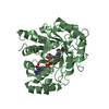
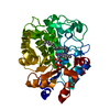
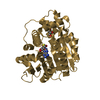
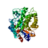
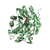
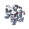
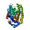
| Title | Multifunctional cellulase, xylanase, mannanase | ||||||
 Components Components | Endoglucanase E | ||||||
 Keywords Keywords | HYDROLASE / cellulase / xylanase / mannanase / multifunction / Endo-1 / 4-beta-glucanase / biomass degradation | ||||||
| Function / homology |  Function and homology information Function and homology informationglucomannan catabolic process / acetylxylan esterase / acetylxylan esterase activity / Hydrolases; Acting on ester bonds; Carboxylic-ester hydrolases / cellulose binding / cellulase / cellulase activity / xylan catabolic process / cellulose catabolic process / extracellular region / metal ion binding Similarity search - Function | ||||||
| Biological species |  Clostridium thermocellum (bacteria) Clostridium thermocellum (bacteria) | ||||||
| Method |  X-RAY DIFFRACTION / SYNCHROTRON / MOLECULAR REPLACEMENT / molecular replacement / Resolution: 2.42 Å X-RAY DIFFRACTION / SYNCHROTRON / MOLECULAR REPLACEMENT / molecular replacement / Resolution: 2.42 Å | ||||||
 Authors Authors | Bianchetti, C.M. / Takasuka, T.E. / Fox, B.G. | ||||||
 Citation Citation | Journal: J.Biol.Chem. / Year: 2020 Title: A structural and kinetic survey of GH5_4 endoglucanases reveals determinants of broad substrate specificity and opportunities for biomass hydrolysis. Authors: Glasgow, E.M. / Kemna, E.I. / Bingman, C.A. / Ing, N. / Deng, K. / Bianchetti, C.M. / Takasuka, T.E. / Northen, T.R. / Fox, B.G. | ||||||
| History |
|
- Structure visualization
| Structure viewer | Molecule: MolmilJmol/JSmol |
|---|
- Downloads & links
-Download
| PDBx/mmCIF format | 4im4.cif.gz | 425.4 KB | Display | PDBx/mmCIF format |
|---|---|---|---|---|
| PDB format | pdb4im4.ent.gz | 348.2 KB | Display | PDB format |
| PDBx/mmJSON format | 4im4.json.gz | Tree view | PDBx/mmJSON format | |
| Others |  Other downloads Other downloads |
-Validation report
| Arichive directory | https://data.pdbj.org/pub/pdb/validation_reports/im/4im4ftp://data.pdbj.org/pub/pdb/validation_reports/im/4im4 | HTTPS FTP |
|---|
-Related structure data
| Related structure data | 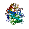 6mq4C 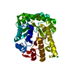 6pz7C 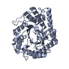 6q1iC 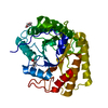 6ui3C 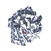 6wqpC  6wqvC 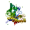 6wqyC 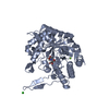 6xrkC 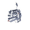 6xsoC 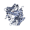 6xsuC  3ndyS  4im1 C: citing same article ( S: Starting model for refinement |
|---|---|
| Similar structure data |


-Links
 PDBj
PDBj- Assembly
| Deposited unit | 
| ||||||||
|---|---|---|---|---|---|---|---|---|---|
| 1 | 
| ||||||||
| 2 | 
| ||||||||
| 3 | 
| ||||||||
| 4 | 
| ||||||||
| 5 | 
| ||||||||
| 6 | 
| ||||||||
| Unit cell |
|
-Components
| #1: Protein | Mass: 38185.832 Da / Num. of mol.: 6 Source method: isolated from a genetically manipulated source Source: (gene. exp.) Clostridium thermocellum (bacteria) / Gene: celE / Plasmid: PVP67K / Production host: #2: Water | ChemComp-HOH / |  Mass: 18.015 Da / Num. of mol.: 1312 / Source method: isolated from a natural source / Formula: H2O Mass: 18.015 Da / Num. of mol.: 1312 / Source method: isolated from a natural source / Formula: H2O |
|---|
-Experimental details
-Experiment
| Experiment | Method: X-RAY DIFFRACTION / Number of used crystals: 1 |
|---|
- Sample preparation
| Crystal | Density Matthews: 2.69 Å3/Da / Density % sol: 54.27 % |
|---|---|
| Crystal grow | Temperature: 298 K / Method: vapor diffusion, hanging drop Details: Protein Solution (20 mg/ml protein, 0.05 M NaCl, 0.010 M MOPS pH 7.0) mixed in a 1:1 ratio with the Well Solution (9% PEG 20,000, 18% mmPEG 550,0.3 M diethyleneglycol, 0.3 M ...Details: Protein Solution (20 mg/ml protein, 0.05 M NaCl, 0.010 M MOPS pH 7.0) mixed in a 1:1 ratio with the Well Solution (9% PEG 20,000, 18% mmPEG 550,0.3 M diethyleneglycol, 0.3 M triethyleneglycol, 0.3 M tetraethyleneglycol, 0.3 M pentaethyleneglycol, 100mM MES/Imidazole buffer pH 6.5). Cryoprotected with 9% PEG 20,000, 18% mmPEG 550, 0.3 M diethyleneglycol, 0.3 M triethyleneglycol, 0.3 M tetraethyleneglycol, 0.3 M pentaethyleneglycol, 100mM MES/Imidazole buffer pH 6.5, 15% ethylene glycol, vapor diffusion, hanging drop, temperature 298K |
-Data collection
| Diffraction | Mean temperature: 100 K | |||||||||||||||||||||||||||||||||||||||||||||||||||||||||||||||||||||||||||||||||||||||||||||||||||||||||||||||||||||||||||||||||||||||||||||||||||
|---|---|---|---|---|---|---|---|---|---|---|---|---|---|---|---|---|---|---|---|---|---|---|---|---|---|---|---|---|---|---|---|---|---|---|---|---|---|---|---|---|---|---|---|---|---|---|---|---|---|---|---|---|---|---|---|---|---|---|---|---|---|---|---|---|---|---|---|---|---|---|---|---|---|---|---|---|---|---|---|---|---|---|---|---|---|---|---|---|---|---|---|---|---|---|---|---|---|---|---|---|---|---|---|---|---|---|---|---|---|---|---|---|---|---|---|---|---|---|---|---|---|---|---|---|---|---|---|---|---|---|---|---|---|---|---|---|---|---|---|---|---|---|---|---|---|---|---|---|
| Diffraction source | Source: SYNCHROTRON / Site: APS  / Beamline: 21-ID-G / Wavelength: 0.97857 Å / Beamline: 21-ID-G / Wavelength: 0.97857 Å | |||||||||||||||||||||||||||||||||||||||||||||||||||||||||||||||||||||||||||||||||||||||||||||||||||||||||||||||||||||||||||||||||||||||||||||||||||
| Detector | Type: MARMOSAIC 300 mm CCD / Detector: CCD / Date: Jun 10, 2012 / Details: mirrors and beryllium lenses | |||||||||||||||||||||||||||||||||||||||||||||||||||||||||||||||||||||||||||||||||||||||||||||||||||||||||||||||||||||||||||||||||||||||||||||||||||
| Radiation | Monochromator: C(111) / Protocol: SINGLE WAVELENGTH / Monochromatic (M) / Laue (L): M / Scattering type: x-ray | |||||||||||||||||||||||||||||||||||||||||||||||||||||||||||||||||||||||||||||||||||||||||||||||||||||||||||||||||||||||||||||||||||||||||||||||||||
| Radiation wavelength | Wavelength: 0.97857 Å / Relative weight: 1 | |||||||||||||||||||||||||||||||||||||||||||||||||||||||||||||||||||||||||||||||||||||||||||||||||||||||||||||||||||||||||||||||||||||||||||||||||||
| Reflection | Resolution: 2.42→50 Å / Num. obs: 92255 / % possible obs: 99.6 % / Redundancy: 4.1 % / Rmerge(I) obs: 0.193 / Χ2: 0.92 / Net I/σ(I): 3.6 | |||||||||||||||||||||||||||||||||||||||||||||||||||||||||||||||||||||||||||||||||||||||||||||||||||||||||||||||||||||||||||||||||||||||||||||||||||
| Reflection shell |
|
-Phasing
| Phasing | Method: molecular replacement | |||||||||
|---|---|---|---|---|---|---|---|---|---|---|
| Phasing MR |
|
- Processing
| Software |
| |||||||||||||||||||||||||||||||||||||||||||||||||||||||||||||||||||||||||||||||||||||||||||||||||||||||||
|---|---|---|---|---|---|---|---|---|---|---|---|---|---|---|---|---|---|---|---|---|---|---|---|---|---|---|---|---|---|---|---|---|---|---|---|---|---|---|---|---|---|---|---|---|---|---|---|---|---|---|---|---|---|---|---|---|---|---|---|---|---|---|---|---|---|---|---|---|---|---|---|---|---|---|---|---|---|---|---|---|---|---|---|---|---|---|---|---|---|---|---|---|---|---|---|---|---|---|---|---|---|---|---|---|---|---|
| Refinement | Method to determine structure: MOLECULAR REPLACEMENT Starting model: PDB entry 3NDY Resolution: 2.42→47.352 Å / Occupancy max: 1 / Occupancy min: 0.13 / SU ML: 0.32 / σ(F): 1.34 / Phase error: 27.09 / Stereochemistry target values: ML
| |||||||||||||||||||||||||||||||||||||||||||||||||||||||||||||||||||||||||||||||||||||||||||||||||||||||||
| Solvent computation | Shrinkage radii: 0.9 Å / VDW probe radii: 1.11 Å / Solvent model: FLAT BULK SOLVENT MODEL | |||||||||||||||||||||||||||||||||||||||||||||||||||||||||||||||||||||||||||||||||||||||||||||||||||||||||
| Displacement parameters | Biso max: 67.2 Å2 / Biso mean: 14.6576 Å2 / Biso min: 1.66 Å2 | |||||||||||||||||||||||||||||||||||||||||||||||||||||||||||||||||||||||||||||||||||||||||||||||||||||||||
| Refinement step | Cycle: LAST / Resolution: 2.42→47.352 Å
| |||||||||||||||||||||||||||||||||||||||||||||||||||||||||||||||||||||||||||||||||||||||||||||||||||||||||
| Refine LS restraints |
| |||||||||||||||||||||||||||||||||||||||||||||||||||||||||||||||||||||||||||||||||||||||||||||||||||||||||
| LS refinement shell | Refine-ID: X-RAY DIFFRACTION / Total num. of bins used: 14
|
