 Movie
Movie Controller


+ Open data

- Basic information
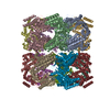
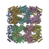
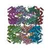
| Entry | Database: PDB / ID: 4hel | ||||||
|---|---|---|---|---|---|---|---|
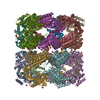
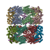
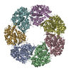
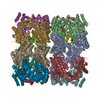
| Title | Crystal structure analysis of apo-GroEL structure | ||||||
 Components Components | 60 kDa chaperonin 4 | ||||||
 Keywords Keywords | CHAPERONE / GroEL / Assist in protein folding / GroES | ||||||
| Function / homology |  Function and homology information Function and homology informationchaperonin ATPase / isomerase activity / ATP-dependent protein folding chaperone / : / protein refolding / ATP binding / metal ion binding / cytoplasm Similarity search - Function | ||||||
| Biological species |  | ||||||
| Method |  X-RAY DIFFRACTION / SYNCHROTRON / MOLECULAR REPLACEMENT / Resolution: 3.2 Å X-RAY DIFFRACTION / SYNCHROTRON / MOLECULAR REPLACEMENT / Resolution: 3.2 Å | ||||||
 Authors Authors | Saxena, A.K. / Meena, S.R. | ||||||
 Citation Citation | Journal: To be Published Title: Crystal structure of apo-GroEL structure Authors: Meena, S.R. / Saxena, A.K. | ||||||
| History |
|
- Structure visualization
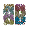
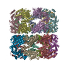
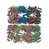
| Structure viewer | Molecule: MolmilJmol/JSmol |
|---|
- Downloads & links
-Download
| PDBx/mmCIF format | 4hel.cif.gz | 1.3 MB | Display | PDBx/mmCIF format |
|---|---|---|---|---|
| PDB format | pdb4hel.ent.gz | 1.1 MB | Display | PDB format |
| PDBx/mmJSON format | 4hel.json.gz | Tree view | PDBx/mmJSON format | |
| Others |  Other downloads Other downloads |
-Validation report
| Arichive directory | https://data.pdbj.org/pub/pdb/validation_reports/he/4helftp://data.pdbj.org/pub/pdb/validation_reports/he/4hel | HTTPS FTP |
|---|
-Related structure data
| Related structure data |  1kp8S S: Starting model for refinement |
|---|---|
| Similar structure data |


-Links
 PDBj
PDBj
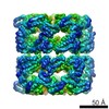
- Assembly
| Deposited unit | 
| ||||||||
|---|---|---|---|---|---|---|---|---|---|
| 1 |
| ||||||||
| Unit cell |
|
-Components
| #1: Protein | Mass: 55349.285 Da / Num. of mol.: 14 / Fragment: GroEL fragment, UNP residues 2-526 / Source method: isolated from a natural source / Source: (natural) #2: Water | ChemComp-HOH / |  Mass: 18.015 Da / Num. of mol.: 901 / Source method: isolated from a natural source / Formula: H2O Mass: 18.015 Da / Num. of mol.: 901 / Source method: isolated from a natural source / Formula: H2O |
|---|
-Experimental details
-Experiment
| Experiment | Method: X-RAY DIFFRACTION / Number of used crystals: 1 |
|---|
- Sample preparation
| Crystal | Density Matthews: 3.25 Å3/Da / Density % sol: 62.14 % |
|---|---|
| Crystal grow | Temperature: 277 K / Method: vapor diffusion, hanging drop / pH: 7.5 Details: 32% MPD, 100mM Tris-HCl, 160mM MgCl2, 10% Glycerol, 2%(w/v) PEG6000, pH 7.5, VAPOR DIFFUSION, HANGING DROP, temperature 277K |
-Data collection
| Diffraction | Mean temperature: 100 K |
|---|---|
| Diffraction source | Source: SYNCHROTRON / Site: ESRF  / Beamline: BM14 / Wavelength: 0.97327 Å / Beamline: BM14 / Wavelength: 0.97327 Å |
| Detector | Type: MARCCD225 / Detector: CCD / Date: Nov 9, 2011 |
| Radiation | Monochromator: crystal / Protocol: SINGLE WAVELENGTH / Monochromatic (M) / Laue (L): M / Scattering type: x-ray |
| Radiation wavelength | Wavelength: 0.97327 Å / Relative weight: 1 |
| Reflection | Resolution: 3.2→59.4 Å / Num. all: 1247571 / Num. obs: 166588 / % possible obs: 100 % / Observed criterion σ(F): 0 / Observed criterion σ(I): 0 / Redundancy: 7.5 % / Biso Wilson estimate: 56.9 Å2 / Rmerge(I) obs: 0.32 / Net I/σ(I): 6.1 |
| Reflection shell | Resolution: 3.2→3.37 Å / Redundancy: 7.3 % / Rmerge(I) obs: 0.85 / Mean I/σ(I) obs: 2.4 / Num. unique all: 24106 / % possible all: 100 |
- Processing
| Software |
| |||||||||||||||||||||||||||||||||||||||||||||||||||||||||||||||||
|---|---|---|---|---|---|---|---|---|---|---|---|---|---|---|---|---|---|---|---|---|---|---|---|---|---|---|---|---|---|---|---|---|---|---|---|---|---|---|---|---|---|---|---|---|---|---|---|---|---|---|---|---|---|---|---|---|---|---|---|---|---|---|---|---|---|---|
| Refinement | Method to determine structure: MOLECULAR REPLACEMENT Starting model: 1KP8 Resolution: 3.2→59.38 Å / Cor.coef. Fo:Fc: 0.913 / Cor.coef. Fo:Fc free: 0.887 / SU B: 16.859 / SU ML: 0.292 / Cross valid method: THROUGHOUT / σ(F): 0 / ESU R Free: 0.429 / Stereochemistry target values: MAXIMUM LIKELIHOOD / Details: HYDROGENS HAVE BEEN ADDED IN THE RIDING POSITIONS
| |||||||||||||||||||||||||||||||||||||||||||||||||||||||||||||||||
| Solvent computation | Ion probe radii: 0.8 Å / Shrinkage radii: 0.8 Å / VDW probe radii: 1.4 Å / Solvent model: MASK | |||||||||||||||||||||||||||||||||||||||||||||||||||||||||||||||||
| Displacement parameters | Biso mean: 43.395 Å2
| |||||||||||||||||||||||||||||||||||||||||||||||||||||||||||||||||
| Refinement step | Cycle: LAST / Resolution: 3.2→59.38 Å
| |||||||||||||||||||||||||||||||||||||||||||||||||||||||||||||||||
| Refine LS restraints |
| |||||||||||||||||||||||||||||||||||||||||||||||||||||||||||||||||
| LS refinement shell | Resolution: 3.2→3.283 Å / Total num. of bins used: 20
|
