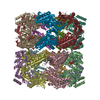
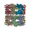
 Movie
Movie Controller

[English] 日本語
 Yorodumi
- PDB-2ynj: GroEL at sub-nanometer resolution by Constrained Single Particle ... -
+ Open data

- Basic information
| Entry | Database: PDB / ID: 2ynj | ||||||
|---|---|---|---|---|---|---|---|
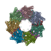
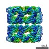
| Title | GroEL at sub-nanometer resolution by Constrained Single Particle Tomography | ||||||
 Components Components | 60 KDA CHAPERONIN | ||||||
 Keywords Keywords | CHAPERONE | ||||||
| Function / homology |  Function and homology information Function and homology informationchaperonin ATPase / isomerase activity / ATP-dependent protein folding chaperone / : / protein refolding / ATP binding / cytoplasm Similarity search - Function | ||||||
| Biological species |  | ||||||
| Method | ELECTRON MICROSCOPY / single particle reconstruction / cryo EM / Resolution: 8.4 Å | ||||||
 Authors Authors | Bartesaghi, A. / Lecumberry, F. / Sapiro, G. / Subramaniam, S. | ||||||
 Citation Citation | Journal: Structure / Year: 2012 Title: Protein secondary structure determination by constrained single-particle cryo-electron tomography. Authors: Alberto Bartesaghi / Federico Lecumberry / Guillermo Sapiro / Sriram Subramaniam /  Abstract: Cryo-electron microscopy (cryo-EM) is a powerful technique for 3D structure determination of protein complexes by averaging information from individual molecular images. The resolutions that can be ...Cryo-electron microscopy (cryo-EM) is a powerful technique for 3D structure determination of protein complexes by averaging information from individual molecular images. The resolutions that can be achieved with single-particle cryo-EM are frequently limited by inaccuracies in assigning molecular orientations based solely on 2D projection images. Tomographic data collection schemes, however, provide powerful constraints that can be used to more accurately determine molecular orientations necessary for 3D reconstruction. Here, we propose "constrained single-particle tomography" as a general strategy for 3D structure determination in cryo-EM. A key component of our approach is the effective use of images recorded in tilt series to extract high-resolution information and correct for the contrast transfer function. By incorporating geometric constraints into the refinement to improve orientational accuracy of images, we reduce model bias and overrefinement artifacts and demonstrate that protein structures can be determined at resolutions of ∼8 Å starting from low-dose tomographic tilt series. | ||||||
| History |
|
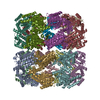
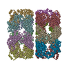
- Structure visualization
| Movie |
Movie viewer |
|---|---|
| Structure viewer | Molecule: MolmilJmol/JSmol |
- Downloads & links
-Download
| PDBx/mmCIF format | 2ynj.cif.gz | 1.1 MB | Display | PDBx/mmCIF format |
|---|---|---|---|---|
| PDB format | pdb2ynj.ent.gz | 922.2 KB | Display | PDB format |
| PDBx/mmJSON format | 2ynj.json.gz | Tree view | PDBx/mmJSON format | |
| Others |  Other downloads Other downloads |
-Validation report
| Arichive directory | https://data.pdbj.org/pub/pdb/validation_reports/yn/2ynjftp://data.pdbj.org/pub/pdb/validation_reports/yn/2ynj | HTTPS FTP |
|---|
-Related structure data
| Related structure data | 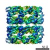 2221MC M: map data used to model this data C: citing same article ( |
|---|---|
| Similar structure data |


-Links
 PDBj
PDBj

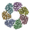
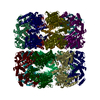
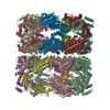
- Assembly
| Deposited unit | 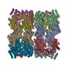
|
|---|---|
| 1 |
|
-Components
| #1: Protein | Mass: 55220.105 Da / Num. of mol.: 14 Source method: isolated from a genetically manipulated source Source: (gene. exp.) #2: Chemical | ChemComp-AGS /   Mass: 523.247 Da / Num. of mol.: 14 / Source method: obtained synthetically / Formula: C10H16N5O12P3S / Comment: ATP-gamma-S, energy-carrying molecule analogue*YM Mass: 523.247 Da / Num. of mol.: 14 / Source method: obtained synthetically / Formula: C10H16N5O12P3S / Comment: ATP-gamma-S, energy-carrying molecule analogue*YM#3: Chemical | ChemComp-TL /   Mass: 204.383 Da / Num. of mol.: 56 / Source method: obtained synthetically / Formula: Tl Mass: 204.383 Da / Num. of mol.: 56 / Source method: obtained synthetically / Formula: Tl#4: Chemical | ChemComp-MG /   Mass: 24.305 Da / Num. of mol.: 14 / Source method: obtained synthetically / Formula: Mg Mass: 24.305 Da / Num. of mol.: 14 / Source method: obtained synthetically / Formula: Mg |
|---|
-Experimental details
-Experiment
| Experiment | Method: ELECTRON MICROSCOPY |
|---|---|
| EM experiment | Aggregation state: PARTICLE / 3D reconstruction method: single particle reconstruction |
- Sample preparation
| Component | Name: GROEL / Type: COMPLEX |
|---|---|
| Buffer solution | pH: 7.5 |
| Specimen | Embedding applied: NO / Shadowing applied: NO / Staining applied: NO / Vitrification applied: YES |
| Specimen support | Details: HOLEY CARBON |
| Vitrification | Instrument: FEI VITROBOT MARK IV / Cryogen name: ETHANE |
- Electron microscopy imaging
| Experimental equipment |  Model: Titan Krios / Image courtesy: FEI Company |
|---|---|
| Microscopy | Model: FEI TITAN KRIOS / Date: Oct 7, 2011 Details: THE TOTAL DOSE OF 25 (ELECTRONS PER SQUARE ANGSTROM) WAS FRACTIONATED EVENLY ACROSS 11 TILTED PROJECTIONS TAKEN BETWEEN 0 AND -20 DEGREES TILT (EVERY 2 DEGREES). |
| Electron gun | Electron source:  FIELD EMISSION GUN / Accelerating voltage: 80 kV / Illumination mode: FLOOD BEAM FIELD EMISSION GUN / Accelerating voltage: 80 kV / Illumination mode: FLOOD BEAM |
| Electron lens | Mode: BRIGHT FIELD / Nominal magnification: 47000 X / Calibrated magnification: 47000 X / Nominal defocus max: 3000 nm / Nominal defocus min: 2000 nm / Cs: 2.7 mm |
| Specimen holder | Temperature: 80 K / Tilt angle max: 0 ° / Tilt angle min: -20 ° |
| Image recording | Electron dose: 25 e/Å2 / Film or detector model: GENERIC CCD |
| Image scans | Num. digital images: 1595 |
| Radiation wavelength | Relative weight: 1 |
- Processing
| EM software |
| ||||||||||||
|---|---|---|---|---|---|---|---|---|---|---|---|---|---|
| CTF correction | Details: DEFOCUS VALUES WERE ASSIGNED TO EACH PARTICLE PROJECTION BASED ON THE DEFOCUS AT THE UNTILTED PLANE OF EACH TILT- SERIES AND A CORRECTION ACCORDING TO THE RELATIVE HEIGHT OF EACH PARTICLE. TO THIS PLANE | ||||||||||||
| Symmetry | Point symmetry: D7 (2x7 fold dihedral) | ||||||||||||
| 3D reconstruction | Method: CONSTRAINED SINGLE PARTICLE TOMOGRAPHY / Resolution: 8.4 Å / Num. of particles: 10000 / Nominal pixel size: 1.74 Å / Actual pixel size: 1.74 Å Details: SUBMISSION BASED ON EXPERIMENTAL DATA FROM EMDB EMD-2221. (DEPOSITION ID: 11174). Symmetry type: POINT | ||||||||||||
| Atomic model building | Protocol: RIGID BODY FIT / Space: REAL / Target criteria: Cross-correlation coefficient / Details: METHOD--RIGID BODY | ||||||||||||
| Atomic model building | PDB-ID: 3.0E+76 / Accession code: 3.0E+76 / Source name: PDB / Type: experimental model | ||||||||||||
| Refinement | Highest resolution: 8.4 Å | ||||||||||||
| Refinement step | Cycle: LAST / Highest resolution: 8.4 Å
|
