 Movie
Movie Controller

[English] 日本語
 Yorodumi
- PDB-4v43: Structural and mechanistic basis for allostery in the bacterial c... -
+ Open data

- Basic information
| Entry | Database: PDB / ID: 4v43 | |||||||||
|---|---|---|---|---|---|---|---|---|---|---|
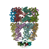
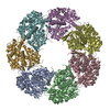
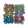
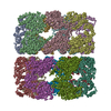
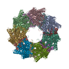
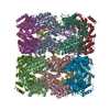
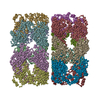
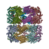
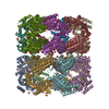
| Title | Structural and mechanistic basis for allostery in the bacterial chaperonin GroEL | |||||||||
 Components Components | GROEL PROTEIN | |||||||||
 Keywords Keywords | CHAPERONE / Wild Type GroEL / ALLOSTERY | |||||||||
| Function / homology |  Function and homology information Function and homology informationGroEL-GroES complex / chaperonin ATPase / virion assembly / isomerase activity / ATP-dependent protein folding chaperone / response to radiation / : / response to heat / protein refolding / protein folding ...GroEL-GroES complex / chaperonin ATPase / virion assembly / isomerase activity / ATP-dependent protein folding chaperone / response to radiation / : / response to heat / protein refolding / protein folding / magnesium ion binding / ATP hydrolysis activity / ATP binding / membrane / identical protein binding / cytosol Similarity search - Function | |||||||||
| Biological species |  | |||||||||
| Method |  X-RAY DIFFRACTION / SYNCHROTRON / MOLECULAR REPLACEMENT / Resolution: 3.52 Å X-RAY DIFFRACTION / SYNCHROTRON / MOLECULAR REPLACEMENT / Resolution: 3.52 Å | |||||||||
 Authors Authors | Wang, J. | |||||||||
 Citation Citation | Journal: To be Published Title: A GroEL/GroES complex structure revisited: the structure-based mechanism of ATP hydrolysis Authors: Wang, J. / Boisvert, D.C. #1: Journal: Nat.Struct.Biol. / Year: 1996Title: The 2.4 A Crystal Structure of the Bacterial Chaperonin GroEL Complexed with ATP Gamma S Authors: Boisvert, D.C. / Wang, J. / Otwinowski, Z. / Horwich, A.L. / Sigler, P.B. #2: Journal: Nature / Year: 1994Title: The Crystal Structure of the Bacterial Chaperonin GroEL at 2.8 A Authors: Braig, K. / Otwinowski, Z. / Hegde, R. / Boisvert, D.C. / Joachimiak, A. / Horwich, A.L. / Sigler, P.B. #3: Journal: Nat.Struct.Biol. / Year: 1995Title: Conformational Variability in the Refined Structure of the Chaperonin GroEL at 2.8 A Resolution Authors: Braig, K. / Adams, P.D. / Brunger, A.T. #4: Journal: Nature / Year: 1997Title: The Crystal Structure of the Asymmetric GroEL-GroES-(ADP)7 Chaperonin Complex Authors: Xu, Z. / Horwich, A.L. / Sigler, P.B. | |||||||||
| History |
|
- Structure visualization
| Structure viewer | Molecule: MolmilJmol/JSmol |
|---|
- Downloads & links
-Download
| PDBx/mmCIF format | 4v43.cif.gz | 2.5 MB | Display | PDBx/mmCIF format |
|---|---|---|---|---|
| PDB format | pdb4v43.ent.gz | Display | PDB format | |
| PDBx/mmJSON format | 4v43.json.gz | Tree view | PDBx/mmJSON format | |
| Others |  Other downloads Other downloads |
-Validation report
| Arichive directory | https://data.pdbj.org/pub/pdb/validation_reports/v4/4v43ftp://data.pdbj.org/pub/pdb/validation_reports/v4/4v43 | HTTPS FTP |
|---|
-Related structure data
| Related structure data |  1kp8S S: Starting model for refinement |
|---|---|
| Similar structure data |

-Links
 PDBj
PDBj
- Assembly
| Deposited unit | 
| ||||||||
|---|---|---|---|---|---|---|---|---|---|
| 1 | 
| ||||||||
| 2 | 
| ||||||||
| Unit cell |
|
-Components
| #1: Protein | Mass: 57158.457 Da / Num. of mol.: 28 / Mutation: D398A Source method: isolated from a genetically manipulated source Source: (gene. exp.) |
|---|
-Experimental details
-Experiment
| Experiment | Method: X-RAY DIFFRACTION / Number of used crystals: 1 |
|---|
- Sample preparation
| Crystal | Density Matthews: 3.35 Å3/Da / Density % sol: 63.25 % |
|---|---|
| Crystal grow | pH: 7 / Details: pH 7.0 |
-Data collection
| Diffraction | Mean temperature: 100 K |
|---|---|
| Diffraction source | Source: SYNCHROTRON / Site: ALS  / Beamline: 5.0.2 / Wavelength: 1 / Beamline: 5.0.2 / Wavelength: 1 |
| Detector | Detector: CCD / Date: Nov 15, 1998 |
| Radiation | Protocol: SINGLE WAVELENGTH / Monochromatic (M) / Laue (L): M / Scattering type: x-ray |
| Radiation wavelength | Wavelength: 1 Å / Relative weight: 1 |
| Reflection | Resolution: 3.5→20 Å / Num. obs: 217987 / % possible obs: 85.1 % / Observed criterion σ(I): -3 / Redundancy: 11.02 % / Rmerge(I) obs: 0.114 |
| Reflection shell | Resolution: 3.5→3.62 Å / Rmerge(I) obs: 0.337 / Mean I/σ(I) obs: 1.396 / % possible all: 51.4 |
- Processing
| Software |
| ||||||||||||||||||||||||||||||||||||||||||||||||||||||||||||
|---|---|---|---|---|---|---|---|---|---|---|---|---|---|---|---|---|---|---|---|---|---|---|---|---|---|---|---|---|---|---|---|---|---|---|---|---|---|---|---|---|---|---|---|---|---|---|---|---|---|---|---|---|---|---|---|---|---|---|---|---|---|
| Refinement | Method to determine structure: MOLECULAR REPLACEMENT Starting model: 1kp8 Resolution: 3.52→20 Å / Rfactor Rfree error: 0.003 / Isotropic thermal model: RESTRAINED / Cross valid method: THROUGHOUT / σ(F): 0
| ||||||||||||||||||||||||||||||||||||||||||||||||||||||||||||
| Solvent computation | Solvent model: FLAT MODEL / Bsol: 11.4049 Å2 / ksol: 0.231794 e/Å3 | ||||||||||||||||||||||||||||||||||||||||||||||||||||||||||||
| Displacement parameters | Biso mean: 80.7 Å2
| ||||||||||||||||||||||||||||||||||||||||||||||||||||||||||||
| Refine analyze |
| ||||||||||||||||||||||||||||||||||||||||||||||||||||||||||||
| Refinement step | Cycle: LAST / Resolution: 3.52→20 Å
| ||||||||||||||||||||||||||||||||||||||||||||||||||||||||||||
| Refine LS restraints |
| ||||||||||||||||||||||||||||||||||||||||||||||||||||||||||||
| LS refinement shell | Resolution: 3.52→3.72 Å / Rfactor Rfree error: 0.015 / Total num. of bins used: 6
| ||||||||||||||||||||||||||||||||||||||||||||||||||||||||||||
| Xplor file |
|
