 Movie
Movie Controller


+ Open data

- Basic information
| Entry | Database: PDB / ID: 1xck | ||||||
|---|---|---|---|---|---|---|---|
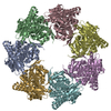
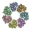
| Title | Crystal structure of apo GroEL | ||||||
 Components Components | 60 kDa chaperonin | ||||||
 Keywords Keywords | CHAPERONE / Chaperonin | ||||||
| Function / homology |  Function and homology information Function and homology informationGroEL-GroES complex / chaperonin ATPase / virion assembly / isomerase activity / ATP-dependent protein folding chaperone / response to radiation / : / protein refolding / response to heat / protein folding ...GroEL-GroES complex / chaperonin ATPase / virion assembly / isomerase activity / ATP-dependent protein folding chaperone / response to radiation / : / protein refolding / response to heat / protein folding / magnesium ion binding / ATP hydrolysis activity / ATP binding / membrane / identical protein binding / cytosol / cytoplasm Similarity search - Function | ||||||
| Biological species |  | ||||||
| Method |  X-RAY DIFFRACTION / SYNCHROTRON / MOLECULAR REPLACEMENT / Resolution: 2.92 Å X-RAY DIFFRACTION / SYNCHROTRON / MOLECULAR REPLACEMENT / Resolution: 2.92 Å | ||||||
 Authors Authors | Bartolucci, C. / Lamba, D. / Grazulis, S. / Manakova, E. / Heumann, H. | ||||||
 Citation Citation | Journal: J.Mol.Biol. / Year: 2005 Title: Crystal structure of wild-type chaperonin GroEL Authors: Bartolucci, C. / Lamba, D. / Grazulis, S. / Manakova, E. / Heumann, H. | ||||||
| History |
|
- Structure visualization
| Structure viewer | Molecule: MolmilJmol/JSmol |
|---|
- Downloads & links
-Download
| PDBx/mmCIF format | 1xck.cif.gz | 1.3 MB | Display | PDBx/mmCIF format |
|---|---|---|---|---|
| PDB format | pdb1xck.ent.gz | 1.1 MB | Display | PDB format |
| PDBx/mmJSON format | 1xck.json.gz | Tree view | PDBx/mmJSON format | |
| Others |  Other downloads Other downloads |
-Validation report
| Arichive directory | https://data.pdbj.org/pub/pdb/validation_reports/xc/1xckftp://data.pdbj.org/pub/pdb/validation_reports/xc/1xck | HTTPS FTP |
|---|
-Related structure data
| Related structure data | 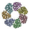 1oelS S: Starting model for refinement |
|---|---|
| Similar structure data |


-Links
 PDBj
PDBj
- Assembly
| Deposited unit | 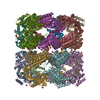
| ||||||||
|---|---|---|---|---|---|---|---|---|---|
| 1 | 
| ||||||||
| 2 | 
| ||||||||
| 3 |
| ||||||||
| Unit cell |
|
-Components
-Protein , 1 types, 14 molecules ABCDEFGHIJKLMN
| #1: Protein | Mass: 57260.504 Da / Num. of mol.: 14 Source method: isolated from a genetically manipulated source Source: (gene. exp.) Production host: Strain (production host): W3110 / References: UniProt: P06139, UniProt: P0A6F5*PLUS |
|---|
-Non-polymers , 5 types, 936 molecules 


| #2: Chemical | ChemComp-SO4 /  Mass: 96.063 Da / Num. of mol.: 43 / Source method: obtained synthetically / Formula: SO4 Mass: 96.063 Da / Num. of mol.: 43 / Source method: obtained synthetically / Formula: SO4#3: Chemical | ChemComp-K /  Mass: 39.098 Da / Num. of mol.: 14 / Source method: obtained synthetically / Formula: K Mass: 39.098 Da / Num. of mol.: 14 / Source method: obtained synthetically / Formula: K#4: Chemical | ChemComp-MPD / (  Mass: 118.174 Da / Num. of mol.: 31 / Source method: obtained synthetically / Formula: C6H14O2 / Comment: precipitant*YM Mass: 118.174 Da / Num. of mol.: 31 / Source method: obtained synthetically / Formula: C6H14O2 / Comment: precipitant*YM#5: Chemical | ChemComp-PEG / |  Mass: 106.120 Da / Num. of mol.: 1 / Source method: obtained synthetically / Formula: C4H10O3 Mass: 106.120 Da / Num. of mol.: 1 / Source method: obtained synthetically / Formula: C4H10O3#6: Water | ChemComp-HOH / | Mass: 18.015 Da / Num. of mol.: 847 / Source method: isolated from a natural source / Formula: H2O |
|---|
-Experimental details
-Experiment
| Experiment | Method: X-RAY DIFFRACTION / Number of used crystals: 1 |
|---|
- Sample preparation
| Crystal | Density Matthews: 3.1 Å3/Da / Density % sol: 60.4 % |
|---|---|
| Crystal grow | Temperature: 291 K / Method: vapor diffusion, sitting drop / pH: 7.5 Details: PEG 4000, ammonium sulphate, hepes, pH 7.5, VAPOR DIFFUSION, SITTING DROP, temperature 291K |
-Data collection
| Diffraction | Mean temperature: 90 K |
|---|---|
| Diffraction source | Source: SYNCHROTRON / Site: MPG/DESY, HAMBURG  / Beamline: BW6 / Wavelength: 1.072 Å / Beamline: BW6 / Wavelength: 1.072 Å |
| Detector | Type: MARRESEARCH / Detector: CCD / Date: Jul 25, 1998 |
| Radiation | Protocol: SINGLE WAVELENGTH / Monochromatic (M) / Laue (L): M / Scattering type: x-ray |
| Radiation wavelength | Wavelength: 1.072 Å / Relative weight: 1 |
| Reflection | Resolution: 2.92→33.71 Å / Num. all: 374890 / Num. obs: 184155 / % possible obs: 83.8 % / Observed criterion σ(F): -3 / Observed criterion σ(I): 0 / Redundancy: 1.7 % / Rsym value: 0.041 / Net I/σ(I): 11.5 |
| Reflection shell | Resolution: 2.92→3 Å / Redundancy: 0.4 % / Mean I/σ(I) obs: 1.5 / Num. unique all: 4965 / Rsym value: 0.158 / % possible all: 27.4 |
- Processing
| Software |
| |||||||||||||||||||||||||
|---|---|---|---|---|---|---|---|---|---|---|---|---|---|---|---|---|---|---|---|---|---|---|---|---|---|---|
| Refinement | Method to determine structure: MOLECULAR REPLACEMENT Starting model: PDB ENTRY 1OEL Resolution: 2.92→33.71 Å / Isotropic thermal model: Restrained / Cross valid method: THROUGHOUT / σ(F): 0 / Stereochemistry target values: Engh & Huber
| |||||||||||||||||||||||||
| Displacement parameters | Biso mean: 48.9 Å2
| |||||||||||||||||||||||||
| Refine analyze |
| |||||||||||||||||||||||||
| Refinement step | Cycle: LAST / Resolution: 2.92→33.71 Å
| |||||||||||||||||||||||||
| Refine LS restraints |
| |||||||||||||||||||||||||
| LS refinement shell | Resolution: 2.9→3.08 Å / Rfactor Rfree error: 0.01
|
