 Movie
Movie Controller


+ Open data

- Basic information
| Entry | Database: PDB / ID: 2eu1 | ||||||
|---|---|---|---|---|---|---|---|
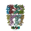
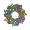
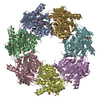
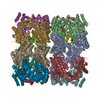
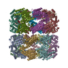
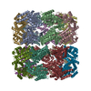
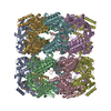
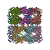
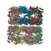
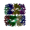
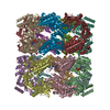
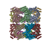
| Title | Crystal structure of the chaperonin GroEL-E461K | ||||||
 Components Components | GROEL | ||||||
 Keywords Keywords | CHAPERONE/PEPTIDE BINDING PROTEIN / chaperonin / GROEL / HSP60 / E461K / CHAPERONE-PEPTIDE BINDING PROTEIN COMPLEX | ||||||
| Function / homology |  Function and homology information Function and homology informationGroEL-GroES complex / chaperonin ATPase / virion assembly / isomerase activity / ATP-dependent protein folding chaperone / response to radiation / : / protein refolding / response to heat / protein folding ...GroEL-GroES complex / chaperonin ATPase / virion assembly / isomerase activity / ATP-dependent protein folding chaperone / response to radiation / : / protein refolding / response to heat / protein folding / magnesium ion binding / ATP hydrolysis activity / ATP binding / membrane / identical protein binding / cytosol Similarity search - Function | ||||||
| Biological species |  | ||||||
| Method |  X-RAY DIFFRACTION / SYNCHROTRON / MOLECULAR REPLACEMENT / Resolution: 3.29 Å X-RAY DIFFRACTION / SYNCHROTRON / MOLECULAR REPLACEMENT / Resolution: 3.29 Å | ||||||
 Authors Authors | Cabo-Bilbao, A. / Spinelli, S. / Sot, B. / Agirre, J. / Mechaly, A.E. / Muga, A. / Guerin, D.M.A. | ||||||
 Citation Citation | Journal: J.Struct.Biol. / Year: 2006 Title: Crystal structure of the temperature-sensitive and allosteric-defective chaperonin GroEL(E461K). Authors: Cabo-Bilbao, A. / Spinelli, S. / Sot, B. / Agirre, J. / Mechaly, A.E. / Muga, A. / Guerin, D.M.A. #1: Journal: J.Mol.Biol. / Year: 2005Title: Crystal structure of wild-type chaperonin GROEL Authors: Bartolucci, C. / Lamba, D. / Grazulis, S. / Manakova, E. / Heumann, H. #2: Journal: Nat Struct Mol Biol / Year: 2004Title: A mutant chaperonin with rearranged inter-ring electrostatic contacts and temperature-sensitive dissociation. Authors: B Trevor Sewell / Robert B Best / Shaoxia Chen / Alan M Roseman / George W Farr / Arthur L Horwich / Helen R Saibil /  Abstract: The chaperonin GroEL assists protein folding through ATP-dependent, cooperative movements that alternately create folding chambers in its two rings. The substitution E461K at the interface between ...The chaperonin GroEL assists protein folding through ATP-dependent, cooperative movements that alternately create folding chambers in its two rings. The substitution E461K at the interface between these two rings causes temperature-sensitive, defective protein folding in Escherichia coli. To understand the molecular defect, we have examined the mutant chaperonin by cryo-EM. The normal out-of-register alignment of contacts between subunits of opposing wild-type rings is changed in E461K to an in-register one. This is associated with loss of cooperativity in ATP binding and hydrolysis. Consistent with the loss of negative cooperativity between rings, the cochaperonin GroES binds simultaneously to both E461K rings. These GroES-bound structures were unstable at higher temperature, dissociating into complexes of single E461K rings associated with GroES. Lacking the allosteric signal from the opposite ring, these complexes cannot release their GroES and become trapped, dead-end states. #3: Journal: Nature / Year: 1995Title: Conformational variability in the refined structure of the chaperonin GroEL at 2.8 A resolution Authors: Braig, K. / Adams, P.D. / Brunger, A.T. #4: Journal: Nature / Year: 1997Title: The crystal structure of the asymmetric GroEL-GroES-(ADP)7 chaperonin complex. Authors: Z Xu / A L Horwich / P B Sigler /  Abstract: Chaperonins assist protein folding with the consumption of ATP. They exist as multi-subunit protein assemblies comprising rings of subunits stacked back to back. In Escherichia coli, asymmetric ...Chaperonins assist protein folding with the consumption of ATP. They exist as multi-subunit protein assemblies comprising rings of subunits stacked back to back. In Escherichia coli, asymmetric intermediates of GroEL are formed with the co-chaperonin GroES and nucleotides bound only to one of the seven-subunit rings (the cis ring) and not to the opposing ring (the trans ring). The structure of the GroEL-GroES-(ADP)7 complex reveals how large en bloc movements of the cis ring's intermediate and apical domains enable bound GroES to stabilize a folding chamber with ADP confined to the cis ring. Elevation and twist of the apical domains double the volume of the central cavity and bury hydrophobic peptide-binding residues in the interface with GroES, as well as between GroEL subunits, leaving a hydrophilic cavity lining that is conducive to protein folding. An inward tilt of the cis equatorial domain causes an outward tilt in the trans ring that opposes the binding of a second GroES. When combined with new functional results, this negative allosteric mechanism suggests a model for an ATP-driven folding cycle that requires a double toroid. #5: Journal: Protein Sci. / Year: 2005 Title: Ionic interactions at both inter-ring contact sites of GroEL are involved in transmission of the allosteric signal: a time-resolved infrared difference study Authors: Sot, B. / von Germar, F. / Mantele, W. / Valpuesta, J.M. / Taneva, S.G. / Muga, A. #6: Journal: J.Biol.Chem. / Year: 2002 Title: Salt bridges at the inter-ring interface regulate the thermostat of GroEL Authors: Sot, B. / Galan, A. / Valpuesta, J.M. / Bertrand, S. / Muga, A. #7: Journal: J.Biol.Chem. / Year: 2003 Title: GroEL stability and function. Contribution of the ionic interactions at the inter-ring contact sites Authors: Sot, B. / Banuelos, S. / Valpuesta, J.M. / Muga, A. | ||||||
| History |
|
- Structure visualization
| Structure viewer | Molecule: MolmilJmol/JSmol |
|---|
- Downloads & links
-Download
| PDBx/mmCIF format | 2eu1.cif.gz | 1.2 MB | Display | PDBx/mmCIF format |
|---|---|---|---|---|
| PDB format | pdb2eu1.ent.gz | 1 MB | Display | PDB format |
| PDBx/mmJSON format | 2eu1.json.gz | Tree view | PDBx/mmJSON format | |
| Others |  Other downloads Other downloads |
-Validation report
| Arichive directory | https://data.pdbj.org/pub/pdb/validation_reports/eu/2eu1ftp://data.pdbj.org/pub/pdb/validation_reports/eu/2eu1 | HTTPS FTP |
|---|
-Related structure data
| Related structure data |  2yeyC 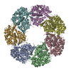 1oelS S: Starting model for refinement C: citing same article ( |
|---|---|
| Similar structure data |

-Links
 PDBj
PDBj
- Assembly
| Deposited unit | 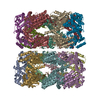
| ||||||||
|---|---|---|---|---|---|---|---|---|---|
| 1 | 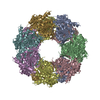
| ||||||||
| 2 | 
| ||||||||
| 3 | 
| ||||||||
| 4 | 
| ||||||||
| Unit cell |
| ||||||||
| Details | The biological GroELE461K molecule is formed in the crystal by the association of two rings from neighbouring asymmetric units. |
-Components
| #1: Protein | Mass: 57391.773 Da / Num. of mol.: 14 / Mutation: E461K Source method: isolated from a genetically manipulated source Source: (gene. exp.) |
|---|
-Experimental details
-Experiment
| Experiment | Method: X-RAY DIFFRACTION / Number of used crystals: 1 |
|---|
- Sample preparation
| Crystal | Density Matthews: 3.11 Å3/Da / Density % sol: 60.41 % |
|---|---|
| Crystal grow | Temperature: 277 K / pH: 8 Details: Reservoir solution: 43% (v/v) MPD, 100mM Imidazole, 170mM MgCl2.6H2O, dissolved in the ratio 1:1.5 with 22.5mg/ml protein, 50mM TRIS-HCL, 10mM MgCl2, pH 8.0, VAPOR DIFFUSION, SITTING DROP, temperature 277K |
-Data collection
| Diffraction | Mean temperature: 100 K |
|---|---|
| Diffraction source | Source: SYNCHROTRON / Site: ESRF  / Beamline: ID29 / Wavelength: 0.97563 / Beamline: ID29 / Wavelength: 0.97563 |
| Radiation | Protocol: SINGLE WAVELENGTH / Monochromatic (M) / Laue (L): M / Scattering type: x-ray |
| Radiation wavelength | Wavelength: 0.97563 Å / Relative weight: 1 |
| Reflection | Resolution: 3.29→39.75 Å / Num. obs: 123350 / % possible obs: 85 % |
- Processing
| Software |
| ||||||||||||||||||||||||||||||||||||||||||||||||||||||||||||
|---|---|---|---|---|---|---|---|---|---|---|---|---|---|---|---|---|---|---|---|---|---|---|---|---|---|---|---|---|---|---|---|---|---|---|---|---|---|---|---|---|---|---|---|---|---|---|---|---|---|---|---|---|---|---|---|---|---|---|---|---|---|
| Refinement | Method to determine structure: MOLECULAR REPLACEMENT Starting model: PDB ENTRY 1OEL Resolution: 3.29→15 Å / σ(F): 0 / Stereochemistry target values: ENGH & HUBER / Details: MAXIMUM LIKELIHOOD TARGET USING AMPLITUDES
| ||||||||||||||||||||||||||||||||||||||||||||||||||||||||||||
| Refinement step | Cycle: LAST / Resolution: 3.29→15 Å
| ||||||||||||||||||||||||||||||||||||||||||||||||||||||||||||
| Refine LS restraints |
| ||||||||||||||||||||||||||||||||||||||||||||||||||||||||||||
| Xplor file | Serial no: 1 / Param file: PROTEIN_REP.PARAM |
