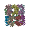
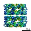
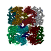
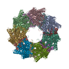
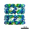
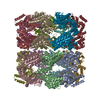
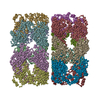
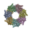
Journal: Nature / Year: 1994 Title: The crystal structure of the bacterial chaperonin GroEL at 2.8 A. Authors: K Braig / Z Otwinowski / R Hegde / D C Boisvert / A Joachimiak / A L Horwich / P B Sigler / Abstract: The crystal structure of Escherichia coli GroEL shows a porous cylinder of 14 subunits made of two nearly 7-fold rotationally symmetrical rings stacked back-to-back with dyad symmetry. The subunits ...The crystal structure of Escherichia coli GroEL shows a porous cylinder of 14 subunits made of two nearly 7-fold rotationally symmetrical rings stacked back-to-back with dyad symmetry. The subunits consist of three domains: a large equatorial domain that forms the foundation of the assembly at its waist and holds the rings together; a large loosely structured apical domain that forms the ends of the cylinder; and a small slender intermediate domain that connects the two, creating side windows. The three-dimensional structure places most of the mutationally defined functional sites on the channel walls and its outward invaginations, and at the ends of the cylinder.
MTRIX THE TRANSFORMATIONS PRESENTED ON MTRIX RECORDS BELOW DESCRIBE NON-CRYSTALLOGRAPHIC RELATIONSHIPS AMONG THE VARIOUS DOMAINS IN THIS ENTRY. APPLYING THE APPROPRIATE MTRIX TRANSFORMATION TO THE RESIDUES LISTED FIRST WILL YIELD APPROXIMATE COORDINATES FOR THE RESIDUES LISTED SECOND. APPLIED TO TRANSFORMED TO MTRIX RESIDUES RESIDUES RMSD M1 6 .. 523 ? 6 .. ? 523 M2 6 .. 523 ? 6 .. ? 523 M3 6 .. 523 ? 6 .. ? 523 M4 6 .. 523 ? 6 .. ? 523 M5 6 .. 523 ? 6 .. ? 523 M6 6 .. 523 ? 6 .. ? 523 THESE TRANSFORMATIONS WILL YIELD APPROXIMATE COORDINATES FOR ONE GROEL 7MER WHEN APPLIED TO THE MONOMER COORDINATES IN THIS ENTRY. THIS STRICT NON-CRYSTALLOGRAPHIC SYMMETRY WAS USED IN THE REFINEMENT.
-
Components
#1: Protein
GROEL (HSP60CLASS)
Mass: 57277.531 Da / Num. of mol.: 7 / Mutation: R13G, A126V Source method: isolated from a genetically manipulated source Source: (gene. exp.) Escherichia coli (E. coli) / Strain: DH5 / Plasmid: IQ-TRC / Gene (production host): GROEL / Production host: Escherichia coli (E. coli) / References: UniProt: P06139, UniProt: P0A6F5*PLUS
-
Experimental details
-
Experiment
Experiment
Method: X-RAY DIFFRACTION
-
Sample preparation
Crystal
Density Matthews: 3.13 Å3/Da / Density % sol: 60.71 %
In the structure databanks used in Yorodumi, some data are registered as the other names, "COVID-19 virus" and "2019-nCoV". Here are the details of the virus and the list of structure data.
Jan 31, 2019. EMDB accession codes are about to change! (news from PDBe EMDB page)
EMDB accession codes are about to change! (news from PDBe EMDB page)
The allocation of 4 digits for EMDB accession codes will soon come to an end. Whilst these codes will remain in use, new EMDB accession codes will include an additional digit and will expand incrementally as the available range of codes is exhausted. The current 4-digit format prefixed with “EMD-” (i.e. EMD-XXXX) will advance to a 5-digit format (i.e. EMD-XXXXX), and so on. It is currently estimated that the 4-digit codes will be depleted around Spring 2019, at which point the 5-digit format will come into force.
The EM Navigator/Yorodumi systems omit the EMD- prefix.
Related info.:Q: What is EMD? / ID/Accession-code notation in Yorodumi/EM Navigator
Yorodumi is a browser for structure data from EMDB, PDB, SASBDB, etc.
This page is also the successor to EM Navigator detail page, and also detail information page/front-end page for Omokage search.
The word "yorodu" (or yorozu) is an old Japanese word meaning "ten thousand". "mi" (miru) is to see.
Related info.:EMDB / PDB / SASBDB / Comparison of 3 databanks / Yorodumi Search / Aug 31, 2016. New EM Navigator & Yorodumi / Yorodumi Papers / Jmol/JSmol / Function and homology information / Changes in new EM Navigator and Yorodumi
 Movie
Movie Controller

 Yorodumi
 Open data
 Basic information
 Components
 Keywords
 Function and homology information
 X-RAY DIFFRACTION / Resolution: 2.8 Å
  Authors
 Citation
 Structure visualization
 Downloads & links
 Other downloads
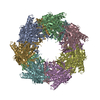
 PDBj
PDBj
 Assembly
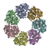
 Sample preparation
 Processing