 Movie
Movie Controller

[English] 日本語
 Yorodumi
- PDB-4f0z: Crystal Structure of Calcineurin in Complex with the Calcineurin-... -
+ Open data

- Basic information
| Entry | Database: PDB / ID: 4f0z | ||||||
|---|---|---|---|---|---|---|---|
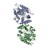
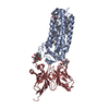
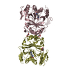
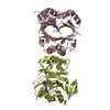
| Title | Crystal Structure of Calcineurin in Complex with the Calcineurin-Inhibiting Domain of the African Swine Fever Virus Protein A238L | ||||||
 Components Components |
| ||||||
 Keywords Keywords | hydrolase/protein binding / EF-hand / phosphatase / PxIxIT / LxVP / calcium signaling / transcription regulation / T-cell activation / Calcineurin inhibition / calmodulin / RCAN / NFAT / heart / nucleus / skeletal muscle / ion channels / hydrolase-protein binding complex | ||||||
| Function / homology |  Function and homology information Function and homology informationnegative regulation of angiotensin-activated signaling pathway / calcium-dependent protein serine/threonine phosphatase regulator activity / regulation of cell proliferation involved in kidney morphogenesis / positive regulation of glomerulus development / negative regulation of calcium ion import across plasma membrane / protein serine/threonine phosphatase complex / negative regulation of signaling / calcium-dependent protein serine/threonine phosphatase activity / positive regulation of saliva secretion / protein serine/threonine phosphatase inhibitor activity ...negative regulation of angiotensin-activated signaling pathway / calcium-dependent protein serine/threonine phosphatase regulator activity / regulation of cell proliferation involved in kidney morphogenesis / positive regulation of glomerulus development / negative regulation of calcium ion import across plasma membrane / protein serine/threonine phosphatase complex / negative regulation of signaling / calcium-dependent protein serine/threonine phosphatase activity / positive regulation of saliva secretion / protein serine/threonine phosphatase inhibitor activity / peptidyl-serine dephosphorylation / calmodulin-dependent protein phosphatase activity / calcineurin complex / positive regulation of connective tissue replacement / positive regulation of calcium ion-dependent exocytosis of neurotransmitter / positive regulation of calcium ion import across plasma membrane / positive regulation of cardiac muscle hypertrophy in response to stress / lung epithelial cell differentiation / negative regulation of dendrite morphogenesis / renal filtration / calcineurin-NFAT signaling cascade / positive regulation of calcineurin-NFAT signaling cascade / myelination in peripheral nervous system / skeletal muscle tissue regeneration / transition between fast and slow fiber / dephosphorylation / positive regulation of osteoclast differentiation / cardiac muscle hypertrophy in response to stress / regulation of synaptic vesicle cycle / positive regulation of activated T cell proliferation / protein dephosphorylation / extrinsic component of plasma membrane / branching involved in blood vessel morphogenesis / dendrite morphogenesis / protein-serine/threonine phosphatase / regulation of postsynaptic neurotransmitter receptor internalization / CLEC7A (Dectin-1) induces NFAT activation / protein serine/threonine phosphatase activity / parallel fiber to Purkinje cell synapse / calcineurin-mediated signaling / epithelial to mesenchymal transition / epidermis development / Calcineurin activates NFAT / Activation of BAD and translocation to mitochondria / DARPP-32 events / positive regulation of osteoblast differentiation / positive regulation of endocytosis / multicellular organismal response to stress / postsynaptic modulation of chemical synaptic transmission / phosphatase binding / keratinocyte differentiation / skeletal muscle fiber development / FCERI mediated Ca+2 mobilization / positive regulation of cell adhesion / T cell activation / hippocampal mossy fiber to CA3 synapse / wound healing / excitatory postsynaptic potential / G1/S transition of mitotic cell cycle / sarcolemma / response to calcium ion / modulation of chemical synaptic transmission / Schaffer collateral - CA1 synapse / Z disc / protein import into nucleus / calcium ion transport / heart development / ATPase binding / Ca2+ pathway / dendritic spine / symbiont-mediated suppression of host NF-kappaB cascade / host cell cytoplasm / calmodulin binding / protein dimerization activity / postsynapse / positive regulation of cell migration / protein domain specific binding / negative regulation of gene expression / calcium ion binding / positive regulation of gene expression / host cell nucleus / glutamatergic synapse / enzyme binding / positive regulation of transcription by RNA polymerase II / mitochondrion / nucleoplasm / plasma membrane / cytoplasm / cytosol Similarity search - Function | ||||||
| Biological species |  Homo sapiens (human) Homo sapiens (human) African swine fever virus Malawi LIL 20/1 African swine fever virus Malawi LIL 20/1 | ||||||
| Method |  X-RAY DIFFRACTION / SYNCHROTRON / MOLECULAR REPLACEMENT / Resolution: 1.7 Å X-RAY DIFFRACTION / SYNCHROTRON / MOLECULAR REPLACEMENT / Resolution: 1.7 Å | ||||||
 Authors Authors | Grigoriu, S. / Peti, W. / Page, R. | ||||||
 Citation Citation | Journal: Plos Biol. / Year: 2013 Title: The molecular mechanism of substrate engagement and immunosuppressant inhibition of calcineurin. Authors: Grigoriu, S. / Bond, R. / Cossio, P. / Chen, J.A. / Ly, N. / Hummer, G. / Page, R. / Cyert, M.S. / Peti, W. | ||||||
| History |
|
- Structure visualization
| Structure viewer | Molecule: MolmilJmol/JSmol |
|---|
- Downloads & links
-Download
| PDBx/mmCIF format | 4f0z.cif.gz | 338.3 KB | Display | PDBx/mmCIF format |
|---|---|---|---|---|
| PDB format | pdb4f0z.ent.gz | 280.8 KB | Display | PDB format |
| PDBx/mmJSON format | 4f0z.json.gz | Tree view | PDBx/mmJSON format | |
| Others |  Other downloads Other downloads |
-Validation report
| Arichive directory | https://data.pdbj.org/pub/pdb/validation_reports/f0/4f0zftp://data.pdbj.org/pub/pdb/validation_reports/f0/4f0z | HTTPS FTP |
|---|
-Related structure data
| Related structure data |  1auiS S: Starting model for refinement |
|---|---|
| Similar structure data |


-Links
 PDBj
PDBj


- Assembly
| Deposited unit | 
| ||||||||
|---|---|---|---|---|---|---|---|---|---|
| 1 |
| ||||||||
| Unit cell |
|
-Components
-Protein , 2 types, 2 molecules AB
| #1: Protein | Mass: 42855.047 Da / Num. of mol.: 1 / Fragment: Calcineurin A Subunit Source method: isolated from a genetically manipulated source Source: (gene. exp.) Homo sapiens (human) / Gene: PPP3CA, CALNA, CNA / Plasmid: p11 / Production host:  References: UniProt: Q08209, protein-serine/threonine phosphatase |
|---|---|
| #2: Protein | Mass: 19322.904 Da / Num. of mol.: 1 / Fragment: Calcineurin B Subunit Source method: isolated from a genetically manipulated source Source: (gene. exp.) Homo sapiens (human) / Gene: PPP3R1, CNA2, CNB / Plasmid: p11 / Production host: References: UniProt: P63098, protein-serine/threonine phosphatase |
-Protein/peptide , 1 types, 1 molecules C
| #3: Protein/peptide | Mass: 5109.087 Da / Num. of mol.: 1 / Fragment: A238L CID (unp residues 200-239) Source method: isolated from a genetically manipulated source Source: (gene. exp.) African swine fever virus Malawi LIL 20/1Strain: Malawi (Lil 20-1) Gene: 5EL, Mal-047, Ordered Locus Name: Mal-047 ORF Name: 5EL Plasmid: pET-RP1B / Production host: |
|---|
-Non-polymers , 3 types, 482 molecules 


| #4: Chemical | ChemComp-GOL /  Mass: 92.094 Da / Num. of mol.: 8 / Source method: obtained synthetically / Formula: C3H8O3 Mass: 92.094 Da / Num. of mol.: 8 / Source method: obtained synthetically / Formula: C3H8O3#5: Chemical | ChemComp-CA /  Mass: 40.078 Da / Num. of mol.: 4 / Source method: obtained synthetically / Formula: Ca Mass: 40.078 Da / Num. of mol.: 4 / Source method: obtained synthetically / Formula: Ca#6: Water | ChemComp-HOH / | Mass: 18.015 Da / Num. of mol.: 470 / Source method: isolated from a natural source / Formula: H2O |
|---|
-Experimental details
-Experiment
| Experiment | Method: X-RAY DIFFRACTION / Number of used crystals: 1 |
|---|
- Sample preparation
| Crystal | Density Matthews: 2.11 Å3/Da / Density % sol: 41.78 % |
|---|---|
| Crystal grow | Temperature: 295 K / Method: vapor diffusion, sitting drop / pH: 5 Details: 0.16 M ammonium citrate dibasic, 20% (w/v) PEG3350, VAPOR DIFFUSION, SITTING DROP, temperature 295K |
-Data collection
| Diffraction | Mean temperature: 100 K |
|---|---|
| Diffraction source | Source: SYNCHROTRON / Site: NSLS  / Beamline: X25 / Wavelength: 1.1 Å / Beamline: X25 / Wavelength: 1.1 Å |
| Detector | Type: PSI PILATUS 6M / Detector: PIXEL / Date: Jun 29, 2011 / Details: Si-111 double crystal monochromator |
| Radiation | Monochromator: Si-111 double crystal monochromator / Protocol: SINGLE WAVELENGTH / Monochromatic (M) / Laue (L): M / Scattering type: x-ray |
| Radiation wavelength | Wavelength: 1.1 Å / Relative weight: 1 |
| Reflection | Resolution: 1.7→50 Å / Num. all: 62191 / Num. obs: 60590 / % possible obs: 97.4 % / Observed criterion σ(I): -3 |
| Reflection shell | Resolution: 1.7→1.73 Å / % possible all: 87.5 |
- Processing
| Software |
| ||||||||||||||||||||||||||||||||||||||||||||||||||||||||||||||||||||||||||||||||||||||||||||||||||||||||||||||||||||||||||||||||||||||||||||||||||||||||||||||||||||||||||||||||||||||||||||||||||||||||||||||||||||||||||||||||||||||||||||||||||||||||||
|---|---|---|---|---|---|---|---|---|---|---|---|---|---|---|---|---|---|---|---|---|---|---|---|---|---|---|---|---|---|---|---|---|---|---|---|---|---|---|---|---|---|---|---|---|---|---|---|---|---|---|---|---|---|---|---|---|---|---|---|---|---|---|---|---|---|---|---|---|---|---|---|---|---|---|---|---|---|---|---|---|---|---|---|---|---|---|---|---|---|---|---|---|---|---|---|---|---|---|---|---|---|---|---|---|---|---|---|---|---|---|---|---|---|---|---|---|---|---|---|---|---|---|---|---|---|---|---|---|---|---|---|---|---|---|---|---|---|---|---|---|---|---|---|---|---|---|---|---|---|---|---|---|---|---|---|---|---|---|---|---|---|---|---|---|---|---|---|---|---|---|---|---|---|---|---|---|---|---|---|---|---|---|---|---|---|---|---|---|---|---|---|---|---|---|---|---|---|---|---|---|---|---|---|---|---|---|---|---|---|---|---|---|---|---|---|---|---|---|---|---|---|---|---|---|---|---|---|---|---|---|---|---|---|---|---|---|---|---|---|---|---|---|---|---|---|---|---|---|---|---|---|
| Refinement | Method to determine structure: MOLECULAR REPLACEMENT Starting model: PDB entry 1AUI Resolution: 1.7→47.313 Å / SU ML: 0.21 / σ(F): 0 / Phase error: 18.7 / Stereochemistry target values: ML
| ||||||||||||||||||||||||||||||||||||||||||||||||||||||||||||||||||||||||||||||||||||||||||||||||||||||||||||||||||||||||||||||||||||||||||||||||||||||||||||||||||||||||||||||||||||||||||||||||||||||||||||||||||||||||||||||||||||||||||||||||||||||||||
| Solvent computation | Shrinkage radii: 0.6 Å / VDW probe radii: 0.9 Å / Solvent model: FLAT BULK SOLVENT MODEL / Bsol: 61.893 Å2 / ksol: 0.433 e/Å3 | ||||||||||||||||||||||||||||||||||||||||||||||||||||||||||||||||||||||||||||||||||||||||||||||||||||||||||||||||||||||||||||||||||||||||||||||||||||||||||||||||||||||||||||||||||||||||||||||||||||||||||||||||||||||||||||||||||||||||||||||||||||||||||
| Displacement parameters |
| ||||||||||||||||||||||||||||||||||||||||||||||||||||||||||||||||||||||||||||||||||||||||||||||||||||||||||||||||||||||||||||||||||||||||||||||||||||||||||||||||||||||||||||||||||||||||||||||||||||||||||||||||||||||||||||||||||||||||||||||||||||||||||
| Refinement step | Cycle: LAST / Resolution: 1.7→47.313 Å
| ||||||||||||||||||||||||||||||||||||||||||||||||||||||||||||||||||||||||||||||||||||||||||||||||||||||||||||||||||||||||||||||||||||||||||||||||||||||||||||||||||||||||||||||||||||||||||||||||||||||||||||||||||||||||||||||||||||||||||||||||||||||||||
| Refine LS restraints |
| ||||||||||||||||||||||||||||||||||||||||||||||||||||||||||||||||||||||||||||||||||||||||||||||||||||||||||||||||||||||||||||||||||||||||||||||||||||||||||||||||||||||||||||||||||||||||||||||||||||||||||||||||||||||||||||||||||||||||||||||||||||||||||
| LS refinement shell |
| ||||||||||||||||||||||||||||||||||||||||||||||||||||||||||||||||||||||||||||||||||||||||||||||||||||||||||||||||||||||||||||||||||||||||||||||||||||||||||||||||||||||||||||||||||||||||||||||||||||||||||||||||||||||||||||||||||||||||||||||||||||||||||
| Refinement TLS params. | Method: refined / Refine-ID: X-RAY DIFFRACTION
| ||||||||||||||||||||||||||||||||||||||||||||||||||||||||||||||||||||||||||||||||||||||||||||||||||||||||||||||||||||||||||||||||||||||||||||||||||||||||||||||||||||||||||||||||||||||||||||||||||||||||||||||||||||||||||||||||||||||||||||||||||||||||||
| Refinement TLS group |
|
