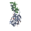
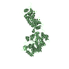
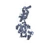
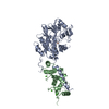
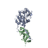
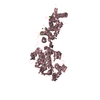
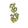
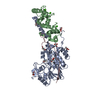
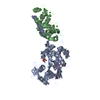
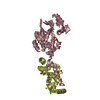
Entry Database : PDB / ID : 3ll8Title Crystal Structure of Calcineurin in Complex with AKAP79 Peptide AKAP79 peptide Calcineurin subunit B type 1 Serine/threonine-protein phosphatase 2B catalytic subunit alpha isoform Keywords / / / / / / / / / / / / / / / / Function / homology Function Domain/homology Component
/ / / / / / / / / / / / / / / / / / / / / / / / / / / / / / / / / / / / / / / / / / / / / / / / / / / / / / / / / / / / / / / / / / / / / / / / / / / / / / / / / / / / / / / / / / / / / / / / / / / / / / / / / / / / / / / / / / / / / / / / / / / / / / / / / / / / / / / / / / / / / / / / / / / Biological species Homo sapiens (human)Method / / / / Resolution : 2 Å Authors Li, H. / Hogan, P.G. #1: Journal : Nature / Year : 1995Title : Crystal structures of human calcineurin and the human FKBP12-FK506-calcineurin complex
Authors: Kissinger, C.R. / Parge, H.E. / Knighton, D.R. / Lewis, C.T. / Pelletier, L.A. / Tempczyk, A. / Kalish, V.J. / Tucker, K.D. / Showalter, R.E. / Moomaw, E.W. / Gastinel, L.N. / Habuka, N. / ... Authors : Kissinger, C.R. / Parge, H.E. / Knighton, D.R. / Lewis, C.T. / Pelletier, L.A. / Tempczyk, A. / Kalish, V.J. / Tucker, K.D. / Showalter, R.E. / Moomaw, E.W. / Gastinel, L.N. / Habuka, N. / Chen, X. / Maldonado, F. / Barker, J.E. / Bacquet, R. / Villafranca, J.E. History Deposition Jan 28, 2010 Deposition site / Processing site Revision 1.0 Jan 12, 2011 Provider / Type Revision 1.1 Jul 13, 2011 Group Revision 1.2 Mar 21, 2012 Group Revision 1.3 Nov 1, 2017 Group / Refinement description / Category / softwareRevision 1.4 Feb 21, 2024 Group Advisory / Data collection ... Advisory / Data collection / Database references / Derived calculations Category chem_comp_atom / chem_comp_bond ... chem_comp_atom / chem_comp_bond / database_2 / pdbx_struct_conn_angle / pdbx_unobs_or_zero_occ_atoms / struct_conn / struct_site Item _database_2.pdbx_DOI / _database_2.pdbx_database_accession ... _database_2.pdbx_DOI / _database_2.pdbx_database_accession / _pdbx_struct_conn_angle.ptnr1_auth_asym_id / _pdbx_struct_conn_angle.ptnr1_auth_comp_id / _pdbx_struct_conn_angle.ptnr1_auth_seq_id / _pdbx_struct_conn_angle.ptnr1_label_asym_id / _pdbx_struct_conn_angle.ptnr1_label_atom_id / _pdbx_struct_conn_angle.ptnr1_label_comp_id / _pdbx_struct_conn_angle.ptnr1_label_seq_id / _pdbx_struct_conn_angle.ptnr2_auth_asym_id / _pdbx_struct_conn_angle.ptnr2_auth_comp_id / _pdbx_struct_conn_angle.ptnr2_auth_seq_id / _pdbx_struct_conn_angle.ptnr2_label_asym_id / _pdbx_struct_conn_angle.ptnr2_label_atom_id / _pdbx_struct_conn_angle.ptnr2_label_comp_id / _pdbx_struct_conn_angle.ptnr3_auth_asym_id / _pdbx_struct_conn_angle.ptnr3_auth_comp_id / _pdbx_struct_conn_angle.ptnr3_auth_seq_id / _pdbx_struct_conn_angle.ptnr3_label_asym_id / _pdbx_struct_conn_angle.ptnr3_label_atom_id / _pdbx_struct_conn_angle.ptnr3_label_comp_id / _pdbx_struct_conn_angle.ptnr3_label_seq_id / _pdbx_struct_conn_angle.value / _struct_conn.pdbx_dist_value / _struct_conn.ptnr1_auth_asym_id / _struct_conn.ptnr1_auth_comp_id / _struct_conn.ptnr1_auth_seq_id / _struct_conn.ptnr1_label_asym_id / _struct_conn.ptnr1_label_atom_id / _struct_conn.ptnr1_label_comp_id / _struct_conn.ptnr1_label_seq_id / _struct_conn.ptnr2_auth_asym_id / _struct_conn.ptnr2_auth_comp_id / _struct_conn.ptnr2_auth_seq_id / _struct_conn.ptnr2_label_asym_id / _struct_conn.ptnr2_label_atom_id / _struct_conn.ptnr2_label_comp_id / _struct_site.pdbx_auth_asym_id / _struct_site.pdbx_auth_comp_id / _struct_site.pdbx_auth_seq_id
Show all Show less
 Movie
Movie Controller


 Open data
 Basic information
 Components
 Keywords
 Function and homology information
 Homo sapiens (human)
 X-RAY DIFFRACTION /
  Authors
 Citation
 Structure visualization
 Downloads & links
 Other downloads
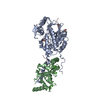

 PDBj
PDBj


 Assembly
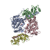


 Mass: 94.971 Da / Num. of mol.: 2 / Source method: obtained synthetically / Formula: PO4
 Mass: 65.409 Da / Num. of mol.: 2 / Source method: obtained synthetically / Formula: Zn
 Mass: 55.845 Da / Num. of mol.: 2 / Source method: obtained synthetically / Formula: Fe
 Mass: 40.078 Da / Num. of mol.: 8 / Source method: obtained synthetically / Formula: Ca
 Sample preparation
 / Beamline: 24-ID-C / Wavelength: 0.9789 Å
 Processing