 Movie
Movie Controller


+ Open data

- Basic information
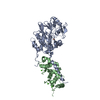
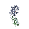
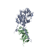
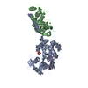
| Entry | Database: PDB / ID: 4il1 | ||||||
|---|---|---|---|---|---|---|---|
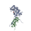
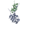
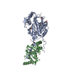
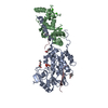
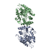
| Title | Crystal Structure of the Rat Calcineurin | ||||||
 Components Components | Calmodulin, Calcineurin subunit B type 1, Serine/threonine-protein phosphatase 2B catalytic subunit alpha isoform | ||||||
 Keywords Keywords | HYDROLASE / Calcium-binding protein / chimera protein / fusion protein / Protein phosphatase | ||||||
| Function / homology |  Function and homology information Function and homology informationActivation of BAD and translocation to mitochondria / Calcineurin activates NFAT / CLEC7A (Dectin-1) induces NFAT activation / histone H2AXS139 phosphatase activity / RNA polymerase II CTD heptapeptide repeat Y1 phosphatase activity / RNA polymerase II CTD heptapeptide repeat T4 phosphatase activity / MAP kinase serine/threonine phosphatase activity / myosin phosphatase activity / FCERI mediated Ca+2 mobilization / negative regulation of angiotensin-activated signaling pathway ...Activation of BAD and translocation to mitochondria / Calcineurin activates NFAT / CLEC7A (Dectin-1) induces NFAT activation / histone H2AXS139 phosphatase activity / RNA polymerase II CTD heptapeptide repeat Y1 phosphatase activity / RNA polymerase II CTD heptapeptide repeat T4 phosphatase activity / MAP kinase serine/threonine phosphatase activity / myosin phosphatase activity / FCERI mediated Ca+2 mobilization / negative regulation of angiotensin-activated signaling pathway / calcium-dependent protein serine/threonine phosphatase regulator activity / regulation of cell proliferation involved in kidney morphogenesis / regulation of store-operated calcium channel activity / positive regulation of glomerulus development / : / negative regulation of signaling / calcium-dependent protein serine/threonine phosphatase activity / positive regulation of saliva secretion / : / calmodulin-dependent protein phosphatase activity / calcineurin complex / slit diaphragm / positive regulation of connective tissue replacement / RNA polymerase II CTD heptapeptide repeat S2 phosphatase activity / RNA polymerase II CTD heptapeptide repeat S7 phosphatase activity / regulation of response to tumor cell / positive regulation of autophagic cell death / DAPK1-calmodulin complex / positive regulation of cardiac muscle hypertrophy in response to stress / lung epithelial cell differentiation / negative regulation of dendrite morphogenesis / renal filtration / RNA polymerase II CTD heptapeptide repeat S5 phosphatase activity / : / : / : / : / : / negative regulation of calcium ion-dependent exocytosis / calcineurin-NFAT signaling cascade / transporter inhibitor activity / myelination in peripheral nervous system / transition between fast and slow fiber / : / type 3 metabotropic glutamate receptor binding / positive regulation of osteoclast differentiation / establishment of protein localization to membrane / Ca2+ pathway / cardiac muscle hypertrophy in response to stress / regulation of synaptic vesicle cycle / positive regulation of activated T cell proliferation / positive regulation of DNA binding / protein dephosphorylation / branching involved in blood vessel morphogenesis / dendrite morphogenesis / negative regulation of high voltage-gated calcium channel activity / protein-serine/threonine phosphatase / regulation of postsynaptic neurotransmitter receptor internalization / response to corticosterone / negative regulation of ryanodine-sensitive calcium-release channel activity / organelle localization by membrane tethering / : / positive regulation of cardiac muscle hypertrophy / autophagosome membrane docking / regulation of synaptic vesicle exocytosis / negative regulation of calcium ion export across plasma membrane / regulation of ryanodine-sensitive calcium-release channel activity / regulation of cardiac muscle cell action potential / presynaptic endocytosis / protein serine/threonine phosphatase activity / parallel fiber to Purkinje cell synapse / calcineurin-mediated signaling / nitric-oxide synthase binding / regulation of cell communication by electrical coupling involved in cardiac conduction / adenylate cyclase binding / protein phosphatase activator activity / epithelial to mesenchymal transition / epidermis development / positive regulation of osteoblast differentiation / regulation of synaptic vesicle endocytosis / positive regulation of endocytosis / detection of calcium ion / multicellular organismal response to stress / postsynaptic cytosol / Schwann cell development / regulation of cardiac muscle contraction / protein localization to nucleus / postsynaptic modulation of chemical synaptic transmission / cell surface receptor signaling pathway via JAK-STAT / catalytic complex / phosphatase binding / keratinocyte differentiation / positive regulation of nitric-oxide synthase activity / phosphatidylinositol 3-kinase binding / activation of adenylate cyclase activity / calcium channel inhibitor activity / presynaptic cytosol / skeletal muscle fiber development / cellular response to interferon-beta / regulation of release of sequestered calcium ion into cytosol by sarcoplasmic reticulum Similarity search - Function | ||||||
| Biological species |  | ||||||
| Method |  X-RAY DIFFRACTION / SYNCHROTRON / MOLECULAR REPLACEMENT / Resolution: 3 Å X-RAY DIFFRACTION / SYNCHROTRON / MOLECULAR REPLACEMENT / Resolution: 3 Å | ||||||
 Authors Authors | Ye, Q. / Faucher, F. / Jia, Z. | ||||||
 Citation Citation | Journal: Cell Signal / Year: 2013 Title: Structural basis of calcineurin activation by calmodulin. Authors: Ye, Q. / Feng, Y. / Yin, Y. / Faucher, F. / Currie, M.A. / Rahman, M.N. / Jin, J. / Li, S. / Wei, Q. / Jia, Z. | ||||||
| History |
|
- Structure visualization
| Structure viewer | Molecule: MolmilJmol/JSmol |
|---|
- Downloads & links
-Download
| PDBx/mmCIF format | 4il1.cif.gz | 448.3 KB | Display | PDBx/mmCIF format |
|---|---|---|---|---|
| PDB format | pdb4il1.ent.gz | 358.6 KB | Display | PDB format |
| PDBx/mmJSON format | 4il1.json.gz | Tree view | PDBx/mmJSON format | |
| Others |  Other downloads Other downloads |
-Validation report
| Arichive directory | https://data.pdbj.org/pub/pdb/validation_reports/il/4il1ftp://data.pdbj.org/pub/pdb/validation_reports/il/4il1 | HTTPS FTP |
|---|
-Related structure data
| Related structure data |  1mf8S S: Starting model for refinement |
|---|---|
| Similar structure data |


-Links
 PDBj
PDBj


- Assembly
| Deposited unit | 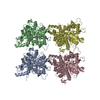
| ||||||||||||||||||||||||||||||||||||||||||||||||||||||||||||||||||||||||||||||||||||||||||||||||||||||||||||||||||||||||||||
|---|---|---|---|---|---|---|---|---|---|---|---|---|---|---|---|---|---|---|---|---|---|---|---|---|---|---|---|---|---|---|---|---|---|---|---|---|---|---|---|---|---|---|---|---|---|---|---|---|---|---|---|---|---|---|---|---|---|---|---|---|---|---|---|---|---|---|---|---|---|---|---|---|---|---|---|---|---|---|---|---|---|---|---|---|---|---|---|---|---|---|---|---|---|---|---|---|---|---|---|---|---|---|---|---|---|---|---|---|---|---|---|---|---|---|---|---|---|---|---|---|---|---|---|---|---|
| 1 | 
| ||||||||||||||||||||||||||||||||||||||||||||||||||||||||||||||||||||||||||||||||||||||||||||||||||||||||||||||||||||||||||||
| 2 | 
| ||||||||||||||||||||||||||||||||||||||||||||||||||||||||||||||||||||||||||||||||||||||||||||||||||||||||||||||||||||||||||||
| 3 | 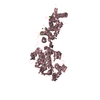
| ||||||||||||||||||||||||||||||||||||||||||||||||||||||||||||||||||||||||||||||||||||||||||||||||||||||||||||||||||||||||||||
| 4 | 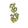
| ||||||||||||||||||||||||||||||||||||||||||||||||||||||||||||||||||||||||||||||||||||||||||||||||||||||||||||||||||||||||||||
| Unit cell |
| ||||||||||||||||||||||||||||||||||||||||||||||||||||||||||||||||||||||||||||||||||||||||||||||||||||||||||||||||||||||||||||
| Noncrystallographic symmetry (NCS) | NCS domain:
NCS domain segments: Component-ID: _ / Beg auth comp-ID: SER / Beg label comp-ID: SER / End auth comp-ID: ARG / End label comp-ID: ARG / Refine code: _
NCS ensembles :
|
-Components
| #1: Protein | Mass: 92698.812 Da / Num. of mol.: 4 Source method: isolated from a genetically manipulated source Source: (gene. exp.) Gene: Calm, Calm1, Calm2, Calm3, Calna, Cam, Cam1, Cam2, Cam3, Camb, Camc, Ppp3ca, Cn a2, Cnb, Ppp3r1 Plasmid: pET21a / Production host:  References: UniProt: P62161, UniProt: P63100, UniProt: P63329, UniProt: P0DP29*PLUS, protein-serine/threonine phosphatase #2: Chemical | ChemComp-CA /   Mass: 40.078 Da / Num. of mol.: 16 / Source method: obtained synthetically / Formula: Ca Mass: 40.078 Da / Num. of mol.: 16 / Source method: obtained synthetically / Formula: Ca#3: Water | ChemComp-HOH / |  Mass: 18.015 Da / Num. of mol.: 124 / Source method: isolated from a natural source / Formula: H2O Mass: 18.015 Da / Num. of mol.: 124 / Source method: isolated from a natural source / Formula: H2OSequence details | THE DEPOSITED PROTEIN SEQUENCE IS A CHIMERA PROTEIN: COMPOSED OF THREE PROTEINS WHICH ARE FUSED BY ...THE DEPOSITED PROTEIN SEQUENCE IS A CHIMERA PROTEIN: COMPOSED OF THREE PROTEINS WHICH ARE FUSED BY TWO GLYCINE LINKERS BETWEEN THEM. THE CLONING CONSTRUCT ORDER FOR THESE THREE PROTEINS IS CALMODULIN | |
|---|
-Experimental details
-Experiment
| Experiment | Method: X-RAY DIFFRACTION / Number of used crystals: 1 |
|---|
- Sample preparation
| Crystal | Density Matthews: 2.8 Å3/Da / Density % sol: 56.05 % |
|---|---|
| Crystal grow | Temperature: 293 K / pH: 5.6 Details: PEG 3350, 1-Propanol, Citric acid, pH 5.6, VAPOR DIFFUSION, HANGING DROP, temperature 293K |
-Data collection
| Diffraction | Mean temperature: 100 K |
|---|---|
| Diffraction source | Source: SYNCHROTRON / Site: CHESS  / Beamline: F1 / Wavelength: 0.917 / Beamline: F1 / Wavelength: 0.917 |
| Detector | Type: ADSC QUANTUM 270 / Detector: CCD / Date: May 6, 2009 / Details: SYNCHROTRON OPTICS |
| Radiation | Monochromator: MONOCHROMATIC MACROMOLECULAR CRYSTALLOGRAPHY / Protocol: SINGLE WAVELENGTH / Monochromatic (M) / Laue (L): M / Scattering type: x-ray |
| Radiation wavelength | Wavelength: 0.917 Å / Relative weight: 1 |
| Reflection | Resolution: 3→10 Å / Num. obs: 81046 / % possible obs: 99.6 % / Observed criterion σ(I): 0 |
| Reflection shell | Resolution: 3→3.1 Å / % possible all: 99.6 |
- Processing
| Software |
| ||||||||||||||||||||||||||||||||||||||||||||||||||||||||||||||||||||||||||||||||||||||||||||||||||||||||||||||||||||||||||||||||||||||||||||||||||||||||||||||||||||||||||||||||||||||
|---|---|---|---|---|---|---|---|---|---|---|---|---|---|---|---|---|---|---|---|---|---|---|---|---|---|---|---|---|---|---|---|---|---|---|---|---|---|---|---|---|---|---|---|---|---|---|---|---|---|---|---|---|---|---|---|---|---|---|---|---|---|---|---|---|---|---|---|---|---|---|---|---|---|---|---|---|---|---|---|---|---|---|---|---|---|---|---|---|---|---|---|---|---|---|---|---|---|---|---|---|---|---|---|---|---|---|---|---|---|---|---|---|---|---|---|---|---|---|---|---|---|---|---|---|---|---|---|---|---|---|---|---|---|---|---|---|---|---|---|---|---|---|---|---|---|---|---|---|---|---|---|---|---|---|---|---|---|---|---|---|---|---|---|---|---|---|---|---|---|---|---|---|---|---|---|---|---|---|---|---|---|---|---|
| Refinement | Method to determine structure: MOLECULAR REPLACEMENT Starting model: 1MF8 Resolution: 3→10 Å / Cor.coef. Fo:Fc: 0.896 / Cor.coef. Fo:Fc free: 0.869 / SU B: 17.535 / SU ML: 0.323 / Cross valid method: THROUGHOUT / σ(F): 0 / ESU R: 0.939 / ESU R Free: 0.385 / Stereochemistry target values: MAXIMUM LIKELIHOOD / Details: HYDROGENS HAVE BEEN ADDED IN THE RIDING POSITIONS
| ||||||||||||||||||||||||||||||||||||||||||||||||||||||||||||||||||||||||||||||||||||||||||||||||||||||||||||||||||||||||||||||||||||||||||||||||||||||||||||||||||||||||||||||||||||||
| Solvent computation | Ion probe radii: 0.8 Å / Shrinkage radii: 0.8 Å / VDW probe radii: 1.2 Å / Solvent model: MASK | ||||||||||||||||||||||||||||||||||||||||||||||||||||||||||||||||||||||||||||||||||||||||||||||||||||||||||||||||||||||||||||||||||||||||||||||||||||||||||||||||||||||||||||||||||||||
| Displacement parameters | Biso mean: 61.91 Å2
| ||||||||||||||||||||||||||||||||||||||||||||||||||||||||||||||||||||||||||||||||||||||||||||||||||||||||||||||||||||||||||||||||||||||||||||||||||||||||||||||||||||||||||||||||||||||
| Refinement step | Cycle: LAST / Resolution: 3→10 Å
| ||||||||||||||||||||||||||||||||||||||||||||||||||||||||||||||||||||||||||||||||||||||||||||||||||||||||||||||||||||||||||||||||||||||||||||||||||||||||||||||||||||||||||||||||||||||
| Refine LS restraints |
|