 Movie
Movie Controller

[English] 日本語
 Yorodumi
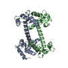
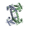
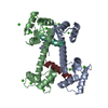
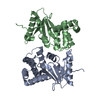
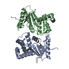
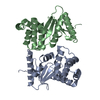
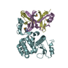
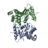
- PDB-2r28: The complex Structure of Calmodulin Bound to a Calcineurin Peptide -
+ Open data

- Basic information
| Entry | Database: PDB / ID: 2r28 | ||||||
|---|---|---|---|---|---|---|---|
| Title | The complex Structure of Calmodulin Bound to a Calcineurin Peptide | ||||||
 Components Components |
| ||||||
 Keywords Keywords | METAL BINDING PROTEIN/Hydrolase / Protein-peptide complex / Acetylation / Calcium / Methylation / Phosphorylation / Ubl conjugation / Alternative splicing / Calmodulin-binding / Hydrolase / Iron / Metal-binding / Nucleus / Protein phosphatase / Zinc / METAL BINDING PROTEIN-Hydrolase COMPLEX | ||||||
| Function / homology |  Function and homology information Function and homology informationnegative regulation of angiotensin-activated signaling pathway / regulation of cell proliferation involved in kidney morphogenesis / positive regulation of glomerulus development / negative regulation of calcium ion import across plasma membrane / negative regulation of signaling / protein serine/threonine phosphatase complex / positive regulation of saliva secretion / peptidyl-serine dephosphorylation / calmodulin-dependent protein phosphatase activity / calcineurin complex ...negative regulation of angiotensin-activated signaling pathway / regulation of cell proliferation involved in kidney morphogenesis / positive regulation of glomerulus development / negative regulation of calcium ion import across plasma membrane / negative regulation of signaling / protein serine/threonine phosphatase complex / positive regulation of saliva secretion / peptidyl-serine dephosphorylation / calmodulin-dependent protein phosphatase activity / calcineurin complex / positive regulation of connective tissue replacement / positive regulation of calcium ion-dependent exocytosis of neurotransmitter / positive regulation of calcium ion import across plasma membrane / positive regulation of cardiac muscle hypertrophy in response to stress / negative regulation of dendrite morphogenesis / renal filtration / : / : / : / : / positive regulation of protein autophosphorylation / : / calcineurin-NFAT signaling cascade / negative regulation of peptidyl-threonine phosphorylation / positive regulation of calcineurin-NFAT signaling cascade / skeletal muscle tissue regeneration / transition between fast and slow fiber / : / dephosphorylation / type 3 metabotropic glutamate receptor binding / positive regulation of osteoclast differentiation / cardiac muscle hypertrophy in response to stress / positive regulation of peptidyl-threonine phosphorylation / positive regulation of activated T cell proliferation / positive regulation of DNA binding / protein dephosphorylation / CaM pathway / extrinsic component of plasma membrane / Cam-PDE 1 activation / Sodium/Calcium exchangers / Calmodulin induced events / Reduction of cytosolic Ca++ levels / Activation of Ca-permeable Kainate Receptor / CREB1 phosphorylation through the activation of CaMKII/CaMKK/CaMKIV cascasde / Loss of phosphorylation of MECP2 at T308 / dendrite morphogenesis / CREB1 phosphorylation through the activation of Adenylate Cyclase / negative regulation of high voltage-gated calcium channel activity / PKA activation / CaMK IV-mediated phosphorylation of CREB / protein-serine/threonine phosphatase / Glycogen breakdown (glycogenolysis) / CLEC7A (Dectin-1) induces NFAT activation / response to corticosterone / negative regulation of ryanodine-sensitive calcium-release channel activity / organelle localization by membrane tethering / Activation of RAC1 downstream of NMDARs / : / autophagosome membrane docking / regulation of synaptic vesicle exocytosis / negative regulation of calcium ion export across plasma membrane / regulation of ryanodine-sensitive calcium-release channel activity / regulation of cardiac muscle cell action potential / presynaptic endocytosis / protein serine/threonine phosphatase activity / Synthesis of IP3 and IP4 in the cytosol / positive regulation of protein serine/threonine kinase activity / Phase 0 - rapid depolarisation / Negative regulation of NMDA receptor-mediated neuronal transmission / Unblocking of NMDA receptors, glutamate binding and activation / RHO GTPases activate PAKs / calcineurin-mediated signaling / nitric-oxide synthase binding / regulation of cell communication by electrical coupling involved in cardiac conduction / Ion transport by P-type ATPases / adenylate cyclase binding / Uptake and function of anthrax toxins / protein phosphatase activator activity / Long-term potentiation / epidermis development / Calcineurin activates NFAT / Regulation of MECP2 expression and activity / DARPP-32 events / Smooth Muscle Contraction / positive regulation of osteoblast differentiation / regulation of synaptic vesicle endocytosis / positive regulation of endocytosis / detection of calcium ion / multicellular organismal response to stress / regulation of cardiac muscle contraction / postsynaptic modulation of chemical synaptic transmission / catalytic complex / keratinocyte differentiation / positive regulation of nitric-oxide synthase activity / RHO GTPases activate IQGAPs / phosphatidylinositol 3-kinase binding / activation of adenylate cyclase activity / calcium channel inhibitor activity / presynaptic cytosol / skeletal muscle fiber development Similarity search - Function | ||||||
| Biological species |  Homo sapiens (human) Homo sapiens (human) | ||||||
| Method |  X-RAY DIFFRACTION / SYNCHROTRON / MOLECULAR REPLACEMENT / Resolution: 1.86 Å X-RAY DIFFRACTION / SYNCHROTRON / MOLECULAR REPLACEMENT / Resolution: 1.86 Å | ||||||
 Authors Authors | Ye, Q. / Zheng, J. / Jia, Z. | ||||||
 Citation Citation | Journal: Proteins / Year: 2008 Title: The complex structure of calmodulin bound to a calcineurin peptide. Authors: Ye, Q. / Wang, H. / Zheng, J. / Wei, Q. / Jia, Z. | ||||||
| History |
|
- Structure visualization
| Structure viewer | Molecule: MolmilJmol/JSmol |
|---|
- Downloads & links
-Download
| PDBx/mmCIF format | 2r28.cif.gz | 84.2 KB | Display | PDBx/mmCIF format |
|---|---|---|---|---|
| PDB format | pdb2r28.ent.gz | 62.6 KB | Display | PDB format |
| PDBx/mmJSON format | 2r28.json.gz | Tree view | PDBx/mmJSON format | |
| Others |  Other downloads Other downloads |
-Validation report
| Arichive directory | https://data.pdbj.org/pub/pdb/validation_reports/r2/2r28ftp://data.pdbj.org/pub/pdb/validation_reports/r2/2r28 | HTTPS FTP |
|---|
-Related structure data
| Related structure data |  2f2oS S: Starting model for refinement |
|---|---|
| Similar structure data |


-Links
 PDBj
PDBj


- Assembly
| Deposited unit | 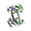
| |||||||||
|---|---|---|---|---|---|---|---|---|---|---|
| 1 |
| |||||||||
| Unit cell |
| |||||||||
| Components on special symmetry positions |
|
-Components
| #1: Protein | Mass: 16852.545 Da / Num. of mol.: 2 / Fragment: Calmodulin-binding domain Source method: isolated from a genetically manipulated source Source: (gene. exp.) Homo sapiens (human) / Strain: Homo sapiens / Gene: CALM1, CALM, CAM, CAM1 / Production host:  #2: Protein/peptide | Mass: 2805.458 Da / Num. of mol.: 2 Source method: isolated from a genetically manipulated source Source: (gene. exp.) Homo sapiens (human) / Strain: Bos taurus / Gene: PPP3CA, CALNA, CNA / Plasmid: pET21a_CciT / Production host: References: UniProt: Q08209, protein-serine/threonine phosphatase #3: Chemical | ChemComp-CA /   Mass: 40.078 Da / Num. of mol.: 8 / Source method: obtained synthetically / Formula: Ca Mass: 40.078 Da / Num. of mol.: 8 / Source method: obtained synthetically / Formula: Ca#4: Water | ChemComp-HOH / |  Mass: 18.015 Da / Num. of mol.: 258 / Source method: isolated from a natural source / Formula: H2O Mass: 18.015 Da / Num. of mol.: 258 / Source method: isolated from a natural source / Formula: H2O |
|---|
-Experimental details
-Experiment
| Experiment | Method: X-RAY DIFFRACTION / Number of used crystals: 1 |
|---|
- Sample preparation
| Crystal | Density Matthews: 2.21 Å3/Da / Density % sol: 44.35 % |
|---|---|
| Crystal grow | Temperature: 298 K / Method: vapor diffusion, hanging drop / pH: 4.5 Details: 20% PEG 3350, 0.2M ammonium phosphate, 0.1M citrite acid , pH 4.5, VAPOR DIFFUSION, HANGING DROP, temperature 298.0K |
-Data collection
| Diffraction | Mean temperature: 100 K |
|---|---|
| Diffraction source | Source: SYNCHROTRON / Site: CHESS  / Beamline: A1 / Wavelength: 0.9124 Å / Beamline: A1 / Wavelength: 0.9124 Å |
| Detector | Type: ADSC QUANTUM 1 / Detector: CCD / Date: Oct 2, 2006 |
| Radiation | Protocol: SINGLE WAVELENGTH / Monochromatic (M) / Laue (L): M / Scattering type: x-ray |
| Radiation wavelength | Wavelength: 0.9124 Å / Relative weight: 1 |
| Reflection | Resolution: 1.85→66.67 Å / Num. all: 29453 / Num. obs: 28898 / % possible obs: 98.1 % / Redundancy: 4.7 % / Rmerge(I) obs: 0.069 / Rsym value: 0.06 / Net I/σ(I): 9 |
| Reflection shell | Resolution: 1.85→1.91 Å / Redundancy: 3.6 % / Rmerge(I) obs: 0.759 / Mean I/σ(I) obs: 8.1 / Num. unique all: 2632 / Rsym value: 0.873 / % possible all: 98.1 |
- Processing
| Software |
| ||||||||||||||||||||
|---|---|---|---|---|---|---|---|---|---|---|---|---|---|---|---|---|---|---|---|---|---|
| Refinement | Method to determine structure: MOLECULAR REPLACEMENT Starting model: PDB entry 2F2O Resolution: 1.86→66.67 Å / Cross valid method: THROUGHOUT / σ(F): 0 / σ(I): 0
| ||||||||||||||||||||
| Refinement step | Cycle: LAST / Resolution: 1.86→66.67 Å
| ||||||||||||||||||||
| LS refinement shell | Resolution: 1.86→1.91 Å / Rfactor Rfree error: 0.17
|
