 Movie
Movie Controller


+ Open data

- Basic information
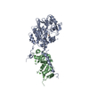
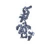
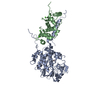
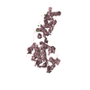
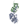
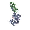
| Entry | Database: PDB / ID: 4orb | ||||||
|---|---|---|---|---|---|---|---|
| Title | Crystal structure of mouse calcineurin | ||||||
 Components Components |
| ||||||
 Keywords Keywords | HYDROLASE/METAL BINDING PROTEIN / Calmodulin-binding / HYDROLASE-METAL BINDING PROTEIN complex | ||||||
| Function / homology |  Function and homology information Function and homology informationActivation of BAD and translocation to mitochondria / positive regulation of angiotensin-activated signaling pathway / negative regulation of angiotensin-activated signaling pathway / regulation of cell proliferation involved in kidney morphogenesis / CLEC7A (Dectin-1) induces NFAT activation / positive regulation of glomerulus development / negative regulation of calcium ion import across plasma membrane / protein serine/threonine phosphatase complex / negative regulation of signaling / calcium-dependent protein serine/threonine phosphatase activity ...Activation of BAD and translocation to mitochondria / positive regulation of angiotensin-activated signaling pathway / negative regulation of angiotensin-activated signaling pathway / regulation of cell proliferation involved in kidney morphogenesis / CLEC7A (Dectin-1) induces NFAT activation / positive regulation of glomerulus development / negative regulation of calcium ion import across plasma membrane / protein serine/threonine phosphatase complex / negative regulation of signaling / calcium-dependent protein serine/threonine phosphatase activity / positive regulation of saliva secretion / Calcineurin activates NFAT / calmodulin-dependent protein phosphatase activity / calcineurin complex / slit diaphragm / positive regulation of connective tissue replacement / positive regulation of calcium ion-dependent exocytosis of neurotransmitter / positive regulation of calcium ion import across plasma membrane / Ca2+ pathway / FCERI mediated Ca+2 mobilization / positive regulation of cardiac muscle hypertrophy in response to stress / lung epithelial cell differentiation / negative regulation of dendrite morphogenesis / renal filtration / renal sodium ion absorption / calcineurin-NFAT signaling cascade / positive regulation of calcineurin-NFAT signaling cascade / myelination in peripheral nervous system / transition between fast and slow fiber / positive regulation of osteoclast differentiation / cardiac muscle hypertrophy in response to stress / regulation of synaptic vesicle cycle / positive regulation of activated T cell proliferation / protein dephosphorylation / branching involved in blood vessel morphogenesis / protein-serine/threonine phosphatase / regulation of postsynaptic neurotransmitter receptor internalization / positive regulation of cardiac muscle hypertrophy / protein serine/threonine phosphatase activity / parallel fiber to Purkinje cell synapse / calcineurin-mediated signaling / epithelial to mesenchymal transition / epidermis development / positive regulation of osteoblast differentiation / positive regulation of endocytosis / multicellular organismal response to stress / Schwann cell development / protein localization to nucleus / postsynaptic modulation of chemical synaptic transmission / phosphatase binding / keratinocyte differentiation / skeletal muscle fiber development / phosphoprotein phosphatase activity / positive regulation of cell adhesion / hippocampal mossy fiber to CA3 synapse / calcium-mediated signaling / cellular response to glucose stimulus / excitatory postsynaptic potential / G1/S transition of mitotic cell cycle / sarcolemma / negative regulation of insulin secretion / response to calcium ion / modulation of chemical synaptic transmission / Schaffer collateral - CA1 synapse / Z disc / cytoplasmic side of plasma membrane / protein import into nucleus / calcium ion transport / heart development / ATPase binding / dendritic spine / proteasome-mediated ubiquitin-dependent protein catabolic process / calmodulin binding / protein dimerization activity / postsynapse / positive regulation of cell migration / protein domain specific binding / negative regulation of gene expression / calcium ion binding / positive regulation of gene expression / synapse / protein-containing complex binding / glutamatergic synapse / enzyme binding / positive regulation of transcription by RNA polymerase II / mitochondrion / metal ion binding / nucleus / plasma membrane / cytoplasm / cytosol Similarity search - Function | ||||||
| Biological species |  | ||||||
| Method |  X-RAY DIFFRACTION / SYNCHROTRON / MOLECULAR REPLACEMENT / Resolution: 3.108 Å X-RAY DIFFRACTION / SYNCHROTRON / MOLECULAR REPLACEMENT / Resolution: 3.108 Å | ||||||
 Authors Authors | Ma, L. / Li, S.J. / Wang, J. / Wu, J.W. / Wang, Z.X. | ||||||
 Citation Citation | Journal: To be Published Title: Cooperative autoinhibition and multi-level activation mechanisms of calcineurin Authors: Li, S.J. / Ma, L. / Wang, J. / Lu, C. / Wang, J. / Wu, J.W. / Wang, Z.X. | ||||||
| History |
|
- Structure visualization
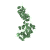
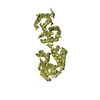
| Structure viewer | Molecule: MolmilJmol/JSmol |
|---|
- Downloads & links
-Download
| PDBx/mmCIF format | 4orb.cif.gz | 237.3 KB | Display | PDBx/mmCIF format |
|---|---|---|---|---|
| PDB format | pdb4orb.ent.gz | 188.9 KB | Display | PDB format |
| PDBx/mmJSON format | 4orb.json.gz | Tree view | PDBx/mmJSON format | |
| Others |  Other downloads Other downloads |
-Validation report
| Arichive directory | https://data.pdbj.org/pub/pdb/validation_reports/or/4orbftp://data.pdbj.org/pub/pdb/validation_reports/or/4orb | HTTPS FTP |
|---|
-Related structure data
| Related structure data |  4or9C  4oraC 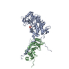 4orcC 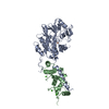 1auiS C: citing same article ( S: Starting model for refinement |
|---|---|
| Similar structure data |


-Links
 PDBj
PDBj


- Assembly
| Deposited unit | 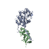
| ||||||||
|---|---|---|---|---|---|---|---|---|---|
| 1 |
| ||||||||
| Unit cell |
|
-Components
-Protein , 2 types, 2 molecules AB
| #1: Protein | Mass: 58754.734 Da / Num. of mol.: 1 / Fragment: catalytic subunit Source method: isolated from a genetically manipulated source Source: (gene. exp.)  References: UniProt: P63328, protein-serine/threonine phosphatase |
|---|---|
| #2: Protein | Mass: 19322.904 Da / Num. of mol.: 1 / Fragment: regulatory subunit Source method: isolated from a genetically manipulated source Source: (gene. exp.) |
-Non-polymers , 4 types, 21 molecules 


| #3: Chemical | ChemComp-ZN /  Mass: 65.409 Da / Num. of mol.: 1 / Source method: obtained synthetically / Formula: Zn Mass: 65.409 Da / Num. of mol.: 1 / Source method: obtained synthetically / Formula: Zn | ||
|---|---|---|---|
| #4: Chemical | ChemComp-FE /  Mass: 55.845 Da / Num. of mol.: 1 / Source method: obtained synthetically / Formula: Fe Mass: 55.845 Da / Num. of mol.: 1 / Source method: obtained synthetically / Formula: Fe | ||
| #5: Chemical | ChemComp-CA /  Mass: 40.078 Da / Num. of mol.: 4 / Source method: obtained synthetically / Formula: Ca Mass: 40.078 Da / Num. of mol.: 4 / Source method: obtained synthetically / Formula: Ca#6: Water | ChemComp-HOH / | Mass: 18.015 Da / Num. of mol.: 15 / Source method: isolated from a natural source / Formula: H2O |
-Details
| Sequence details | AMINO ACIDS (447-456) ARE MISSING BECAUSE THIS SEQUENCE CORRESPOND |
|---|
-Experimental details
-Experiment
| Experiment | Method: X-RAY DIFFRACTION / Number of used crystals: 1 |
|---|
- Sample preparation
| Crystal | Density Matthews: 2.96 Å3/Da / Density % sol: 58.39 % |
|---|---|
| Crystal grow | Temperature: 294 K / Method: vapor diffusion, hanging drop / pH: 6.1 Details: 100mM MES, pH 6.1, 18% PEG3350, 8% Glycerol, VAPOR DIFFUSION, HANGING DROP, temperature 294K |
-Data collection
| Diffraction | Mean temperature: 100 K |
|---|---|
| Diffraction source | Source: SYNCHROTRON / Site: SPring-8  / Beamline: BL41XU / Wavelength: 1 Å / Beamline: BL41XU / Wavelength: 1 Å |
| Detector | Type: RAYONIX MX225HE / Detector: CCD / Date: Jul 21, 2010 |
| Radiation | Monochromator: rotated-inclined double-crystal monochromator Protocol: SINGLE WAVELENGTH / Monochromatic (M) / Laue (L): M / Scattering type: x-ray |
| Radiation wavelength | Wavelength: 1 Å / Relative weight: 1 |
| Reflection | Resolution: 3.1→40 Å / Num. all: 17090 / Num. obs: 17038 / % possible obs: 98 % / Observed criterion σ(F): 1 / Observed criterion σ(I): 1 / Redundancy: 4.3 % / Biso Wilson estimate: 61.1 Å2 / Rmerge(I) obs: 0.12 / Net I/σ(I): 11.4 |
| Reflection shell | Resolution: 3.1→3.21 Å / Redundancy: 4 % / Rmerge(I) obs: 0.606 / Mean I/σ(I) obs: 2.273 / Num. unique all: 1587 / % possible all: 92.8 |
- Processing
| Software |
| ||||||||||||||||||||||||||||||||||||||||||||||||||||||||||||||||||||||||||||||||||||||||||||||||||||
|---|---|---|---|---|---|---|---|---|---|---|---|---|---|---|---|---|---|---|---|---|---|---|---|---|---|---|---|---|---|---|---|---|---|---|---|---|---|---|---|---|---|---|---|---|---|---|---|---|---|---|---|---|---|---|---|---|---|---|---|---|---|---|---|---|---|---|---|---|---|---|---|---|---|---|---|---|---|---|---|---|---|---|---|---|---|---|---|---|---|---|---|---|---|---|---|---|---|---|---|---|---|
| Refinement | Method to determine structure: MOLECULAR REPLACEMENT Starting model: 1AUI Resolution: 3.108→39.691 Å / SU ML: 0.42 / σ(F): 1.35 / Phase error: 22.86 / Stereochemistry target values: ML
| ||||||||||||||||||||||||||||||||||||||||||||||||||||||||||||||||||||||||||||||||||||||||||||||||||||
| Solvent computation | Shrinkage radii: 0.9 Å / VDW probe radii: 1.11 Å / Solvent model: FLAT BULK SOLVENT MODEL / Bsol: 37.512 Å2 / ksol: 0.377 e/Å3 | ||||||||||||||||||||||||||||||||||||||||||||||||||||||||||||||||||||||||||||||||||||||||||||||||||||
| Displacement parameters |
| ||||||||||||||||||||||||||||||||||||||||||||||||||||||||||||||||||||||||||||||||||||||||||||||||||||
| Refinement step | Cycle: LAST / Resolution: 3.108→39.691 Å
| ||||||||||||||||||||||||||||||||||||||||||||||||||||||||||||||||||||||||||||||||||||||||||||||||||||
| Refine LS restraints |
| ||||||||||||||||||||||||||||||||||||||||||||||||||||||||||||||||||||||||||||||||||||||||||||||||||||
| LS refinement shell | Refine-ID: X-RAY DIFFRACTION / Total num. of bins used: 6
| ||||||||||||||||||||||||||||||||||||||||||||||||||||||||||||||||||||||||||||||||||||||||||||||||||||
| Refinement TLS params. | Method: refined / Refine-ID: X-RAY DIFFRACTION
| ||||||||||||||||||||||||||||||||||||||||||||||||||||||||||||||||||||||||||||||||||||||||||||||||||||
| Refinement TLS group |
|
