 Movie
Movie Controller

[English] 日本語
 Yorodumi
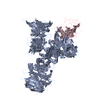
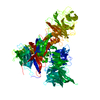
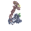
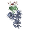
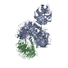
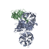
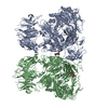
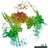
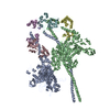
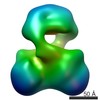
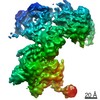
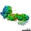
- PDB-4a0l: Structure of DDB1-DDB2-CUL4B-RBX1 bound to a 12 bp abasic site co... -
+ Open data

- Basic information
| Entry | Database: PDB / ID: 4a0l | ||||||
|---|---|---|---|---|---|---|---|
| Title | Structure of DDB1-DDB2-CUL4B-RBX1 bound to a 12 bp abasic site containing DNA-duplex | ||||||
 Components Components |
| ||||||
 Keywords Keywords | LIGASE/DNA-BINDING PROTEIN/DNA / LIGASE-DNA-BINDING PROTEIN-DNA COMPLEX | ||||||
| Function / homology |  Function and homology information Function and homology informationDual Incision in GG-NER / DNA Damage Recognition in GG-NER / Formation of Incision Complex in GG-NER / Neddylation / Prolactin receptor signaling / Recognition of DNA damage by PCNA-containing replication complex / Formation of TC-NER Pre-Incision Complex / DNA Damage Recognition in GG-NER / Dual Incision in GG-NER / Dual incision in TC-NER ...Dual Incision in GG-NER / DNA Damage Recognition in GG-NER / Formation of Incision Complex in GG-NER / Neddylation / Prolactin receptor signaling / Recognition of DNA damage by PCNA-containing replication complex / Formation of TC-NER Pre-Incision Complex / DNA Damage Recognition in GG-NER / Dual Incision in GG-NER / Dual incision in TC-NER / Gap-filling DNA repair synthesis and ligation in TC-NER / Formation of Incision Complex in GG-NER / Regulation of BACH1 activity / Regulation of RAS by GAPs / Degradation of DVL / Ubiquitin-Mediated Degradation of Phosphorylated Cdc25A / Regulation of RUNX2 expression and activity / Degradation of GLI1 by the proteasome / Hedgehog 'on' state / FBXL7 down-regulates AURKA during mitotic entry and in early mitosis / Orc1 removal from chromatin / GSK3B and BTRC:CUL1-mediated-degradation of NFE2L2 / eukaryotic initiation factor 4E binding / Oxygen-dependent proline hydroxylation of Hypoxia-inducible Factor Alpha / Degradation of beta-catenin by the destruction complex / base-excision repair, AP site formation via deaminated base removal / miRNA-mediated gene silencing by mRNA destabilization / anaphase-promoting complex / Interleukin-1 signaling / GLI3 is processed to GLI3R by the proteasome / Neddylation / cullin-RING-type E3 NEDD8 transferase / KEAP1-NFE2L2 pathway / astrocyte differentiation / cullin-RING ubiquitin ligase complex / Cul7-RING ubiquitin ligase complex / Antigen processing: Ubiquitination & Proteasome degradation / positive regulation of protein autoubiquitination / positive regulation by virus of viral protein levels in host cell / RNA polymerase II transcription initiation surveillance / protein neddylation / positive regulation of epithelial cell apoptotic process / spindle assembly involved in female meiosis / NEDD8 ligase activity / epigenetic programming in the zygotic pronuclei / negative regulation of response to oxidative stress / VCB complex / Cul5-RING ubiquitin ligase complex / UV-damage excision repair / ubiquitin-ubiquitin ligase activity / ubiquitin-dependent protein catabolic process via the C-end degron rule pathway / SCF ubiquitin ligase complex / lysosome organization / Cul2-RING ubiquitin ligase complex / biological process involved in interaction with symbiont / type I interferon-mediated signaling pathway / Cul3-RING ubiquitin ligase complex / regulation of mitotic cell cycle phase transition / negative regulation of type I interferon production / WD40-repeat domain binding / p38MAPK cascade / SCF-dependent proteasomal ubiquitin-dependent protein catabolic process / Cul4A-RING E3 ubiquitin ligase complex / Cul4-RING E3 ubiquitin ligase complex / Cul4B-RING E3 ubiquitin ligase complex / negative regulation of signal transduction by p53 class mediator / ubiquitin ligase complex scaffold activity / negative regulation of reproductive process / negative regulation of developmental process / TORC1 signaling / epithelial to mesenchymal transition / viral release from host cell / cullin family protein binding / protein monoubiquitination / ectopic germ cell programmed cell death / negative regulation of ubiquitin-dependent protein catabolic process / response to UV / ubiquitin ligase complex / positive regulation of G1/S transition of mitotic cell cycle / positive regulation of viral genome replication / autophagosome assembly / mitophagy / site of DNA damage / positive regulation of type I interferon production / signal transduction in response to DNA damage / negative regulation of TORC1 signaling / protein K48-linked ubiquitination / negative regulation of insulin receptor signaling pathway / proteasomal protein catabolic process / positive regulation of TORC1 signaling / transcription-coupled nucleotide-excision repair / negative regulation of proteasomal ubiquitin-dependent protein catabolic process / positive regulation of autophagy / regulation of mitotic cell cycle / cellular response to nutrient levels / positive regulation of gluconeogenesis / cellular response to amino acid starvation / T cell activation / negative regulation of autophagy / protein catabolic process Similarity search - Function | ||||||
| Biological species |  HOMO SAPIENS (human) HOMO SAPIENS (human)  SYNTHETIC CONSTRUCT (others) | ||||||
| Method |  X-RAY DIFFRACTION / SYNCHROTRON / MOLECULAR REPLACEMENT / Resolution: 7.4 Å X-RAY DIFFRACTION / SYNCHROTRON / MOLECULAR REPLACEMENT / Resolution: 7.4 Å | ||||||
 Authors Authors | Fischer, E.S. / Scrima, A. / Gut, H. / Thoma, N.H. | ||||||
 Citation Citation | Journal: Cell(Cambridge,Mass.) / Year: 2011 Title: The Molecular Basis of Crl4(Ddb2/Csa) Ubiquitin Ligase Architecture, Targeting, and Activation. Authors: Fischer, E.S. / Scrima, A. / Bohm, K. / Matsumoto, S. / Lingaraju, G.M. / Faty, M. / Yasuda, T. / Cavadini, S. / Wakasugi, M. / Hanaoka, F. / Iwai, S. / Gut, H. / Sugasawa, K. / Thoma, N.H. | ||||||
| History |
|
- Structure visualization
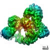
| Structure viewer | Molecule: MolmilJmol/JSmol |
|---|
- Downloads & links
-Download
| PDBx/mmCIF format | 4a0l.cif.gz | 1.8 MB | Display | PDBx/mmCIF format |
|---|---|---|---|---|
| PDB format | pdb4a0l.ent.gz | 1.5 MB | Display | PDB format |
| PDBx/mmJSON format | 4a0l.json.gz | Tree view | PDBx/mmJSON format | |
| Others |  Other downloads Other downloads |
-Validation report
| Arichive directory | https://data.pdbj.org/pub/pdb/validation_reports/a0/4a0lftp://data.pdbj.org/pub/pdb/validation_reports/a0/4a0l | HTTPS FTP |
|---|
-Related structure data
| Related structure data | 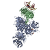 4a08C 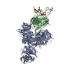 4a09C 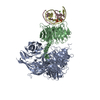 4a0aC 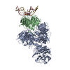 4a0bC 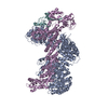 4a0cSC 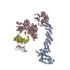 4a0kC  4a11C 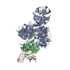 3ei2S C: citing same article ( S: Starting model for refinement |
|---|---|
| Similar structure data |

-Links
 PDBj
PDBj


- Assembly
| Deposited unit | 
| ||||||||
|---|---|---|---|---|---|---|---|---|---|
| 1 | 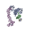
| ||||||||
| 2 | 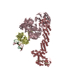
| ||||||||
| Unit cell |
|
-Components
-DNA DAMAGE-BINDING PROTEIN ... , 2 types, 4 molecules ACBD
| #1: Protein | Mass: 127425.812 Da / Num. of mol.: 2 Source method: isolated from a genetically manipulated source Source: (gene. exp.) HOMO SAPIENS (human) / Plasmid: PFASTBAC DERIVED / Cell line (production host): High Five / Production host:  TRICHOPLUSIA NI (cabbage looper) / References: UniProt: Q16531 TRICHOPLUSIA NI (cabbage looper) / References: UniProt: Q16531#2: Protein | Mass: 43418.102 Da / Num. of mol.: 2 / Fragment: RESIDUES 60-423 Source method: isolated from a genetically manipulated source Details: VARIANT WITH GLN AT POSITION 180 AND ARG AT POSITION 214 SIMILAR TO PDB ENTRY 3EI2 Source: (gene. exp.) TRICHOPLUSIA NI (cabbage looper) / References: UniProt: Q2YDS1 |
|---|
-Protein , 2 types, 4 molecules EHFI
| #3: Protein | Mass: 84992.195 Da / Num. of mol.: 2 / Fragment: RESIDUES 193-913 Source method: isolated from a genetically manipulated source Source: (gene. exp.) HOMO SAPIENS (human) / Plasmid: PFASTBAC DERIVED / Cell line (production host): High Five / Production host: TRICHOPLUSIA NI (cabbage looper) / References: UniProt: Q13620#4: Protein | Mass: 11330.942 Da / Num. of mol.: 2 / Fragment: RESIDUES 12-108 Source method: isolated from a genetically manipulated source Source: (gene. exp.) TRICHOPLUSIA NI (cabbage looper) / References: UniProt: P62878 |
|---|
-DNA chain , 2 types, 4 molecules RTSU
| #5: DNA chain | Mass: 3498.283 Da / Num. of mol.: 2 / Source method: obtained synthetically / Source: (synth.) SYNTHETIC CONSTRUCT (others) #6: DNA chain | Mass: 3702.428 Da / Num. of mol.: 2 / Source method: obtained synthetically / Source: (synth.) SYNTHETIC CONSTRUCT (others) |
|---|
-Experimental details
-Experiment
| Experiment | Method: X-RAY DIFFRACTION / Number of used crystals: 1 |
|---|
- Sample preparation
| Crystal | Density Matthews: 4.6 Å3/Da / Density % sol: 73 % / Description: NONE |
|---|---|
| Crystal grow | pH: 6.2 / Details: 100MM MES PH 6.2, 3.1% PEG 6000, 4% ETHYLENEGLYCOL |
-Data collection
| Diffraction | Mean temperature: 100 K |
|---|---|
| Diffraction source | Source: SYNCHROTRON / Site: SLS  / Beamline: X06DA / Wavelength: 1.0015 / Beamline: X06DA / Wavelength: 1.0015 |
| Detector | Type: MARMOSAIC 225 mm CCD / Detector: CCD / Date: Aug 17, 2009 |
| Radiation | Protocol: SINGLE WAVELENGTH / Monochromatic (M) / Laue (L): M / Scattering type: x-ray |
| Radiation wavelength | Wavelength: 1.0015 Å / Relative weight: 1 |
| Reflection | Resolution: 7.4→30 Å / Num. obs: 13547 / % possible obs: 97.5 % / Observed criterion σ(I): -3 / Redundancy: 3.7 % / Rmerge(I) obs: 0.09 / Net I/σ(I): 10.2 |
| Reflection shell | Resolution: 7.4→7.59 Å / Redundancy: 3.7 % / Rmerge(I) obs: 0.62 / Mean I/σ(I) obs: 2.3 / % possible all: 99.1 |
- Processing
| Software |
| |||||||||||||||||||||||||||||||||||||||||||||||||||||||||||||||||||||||||||||||||||||||||||||||||||||||||||||||||||||||||||||||||||||||||||||||||||||||||||||||||||||||||||||||
|---|---|---|---|---|---|---|---|---|---|---|---|---|---|---|---|---|---|---|---|---|---|---|---|---|---|---|---|---|---|---|---|---|---|---|---|---|---|---|---|---|---|---|---|---|---|---|---|---|---|---|---|---|---|---|---|---|---|---|---|---|---|---|---|---|---|---|---|---|---|---|---|---|---|---|---|---|---|---|---|---|---|---|---|---|---|---|---|---|---|---|---|---|---|---|---|---|---|---|---|---|---|---|---|---|---|---|---|---|---|---|---|---|---|---|---|---|---|---|---|---|---|---|---|---|---|---|---|---|---|---|---|---|---|---|---|---|---|---|---|---|---|---|---|---|---|---|---|---|---|---|---|---|---|---|---|---|---|---|---|---|---|---|---|---|---|---|---|---|---|---|---|---|---|---|---|---|
| Refinement | Method to determine structure: MOLECULAR REPLACEMENT Starting model: PDB ENTRIES 3EI2, 4A0C Resolution: 7.4→29.862 Å / SU ML: 2.21 / σ(F): 2 / Phase error: 34.97 / Stereochemistry target values: ML Details: THE MOLECULAR REPLACEMENT SOLUTION HAS BEEN RIGID BODY REFINED TO OBTAIN THE OVERALL ASSEMBLY OF THE COMPLEX. NO REBUILDING HAS BEEN PERFORMED DUE TO LIMITED RESOLUTION. RBX1 RESIDUES 40-108 ...Details: THE MOLECULAR REPLACEMENT SOLUTION HAS BEEN RIGID BODY REFINED TO OBTAIN THE OVERALL ASSEMBLY OF THE COMPLEX. NO REBUILDING HAS BEEN PERFORMED DUE TO LIMITED RESOLUTION. RBX1 RESIDUES 40-108 AND CUL4B RESIDUES 208-209 HAVE BEEN REMOVED DUE TO UNCERTAINTY OF CONFORMATIONS. STEREOCHEMISTRY IS BASED ON THE SEARCH MODELS 3EI2 AND 4A0C.
| |||||||||||||||||||||||||||||||||||||||||||||||||||||||||||||||||||||||||||||||||||||||||||||||||||||||||||||||||||||||||||||||||||||||||||||||||||||||||||||||||||||||||||||||
| Solvent computation | Shrinkage radii: 0.9 Å / VDW probe radii: 1.11 Å / Solvent model: FLAT BULK SOLVENT MODEL / Bsol: 143.782 Å2 / ksol: 0.313 e/Å3 | |||||||||||||||||||||||||||||||||||||||||||||||||||||||||||||||||||||||||||||||||||||||||||||||||||||||||||||||||||||||||||||||||||||||||||||||||||||||||||||||||||||||||||||||
| Displacement parameters | Biso mean: 344 Å2 | |||||||||||||||||||||||||||||||||||||||||||||||||||||||||||||||||||||||||||||||||||||||||||||||||||||||||||||||||||||||||||||||||||||||||||||||||||||||||||||||||||||||||||||||
| Refinement step | Cycle: LAST / Resolution: 7.4→29.862 Å
| |||||||||||||||||||||||||||||||||||||||||||||||||||||||||||||||||||||||||||||||||||||||||||||||||||||||||||||||||||||||||||||||||||||||||||||||||||||||||||||||||||||||||||||||
| Refine LS restraints |
| |||||||||||||||||||||||||||||||||||||||||||||||||||||||||||||||||||||||||||||||||||||||||||||||||||||||||||||||||||||||||||||||||||||||||||||||||||||||||||||||||||||||||||||||
| LS refinement shell |
| |||||||||||||||||||||||||||||||||||||||||||||||||||||||||||||||||||||||||||||||||||||||||||||||||||||||||||||||||||||||||||||||||||||||||||||||||||||||||||||||||||||||||||||||
| Refinement TLS params. | Method: refined / Refine-ID: X-RAY DIFFRACTION
| |||||||||||||||||||||||||||||||||||||||||||||||||||||||||||||||||||||||||||||||||||||||||||||||||||||||||||||||||||||||||||||||||||||||||||||||||||||||||||||||||||||||||||||||
| Refinement TLS group |
|
