 Movie
Movie Controller


+ Open data

- Basic information
| Entry | Database: PDB / ID: 2hye | ||||||
|---|---|---|---|---|---|---|---|
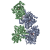
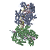
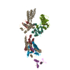
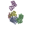
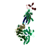
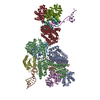
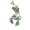
| Title | Crystal Structure of the DDB1-Cul4A-Rbx1-SV5V Complex | ||||||
 Components Components |
| ||||||
 Keywords Keywords | PROTEIN BINDING / beta propeller / RING finger / zinc finger / propeller cluster / helical repeats / cullin repeats | ||||||
| Function / homology |  Function and homology information Function and homology informationnegative regulation of granulocyte differentiation / negative regulation of beige fat cell differentiation / cullin-RING-type E3 NEDD8 transferase / NEDD8 transferase activity / cullin-RING ubiquitin ligase complex / Cul7-RING ubiquitin ligase complex / cellular response to chemical stress / Loss of Function of FBXW7 in Cancer and NOTCH1 Signaling / regulation of DNA damage checkpoint / positive regulation of protein autoubiquitination ...negative regulation of granulocyte differentiation / negative regulation of beige fat cell differentiation / cullin-RING-type E3 NEDD8 transferase / NEDD8 transferase activity / cullin-RING ubiquitin ligase complex / Cul7-RING ubiquitin ligase complex / cellular response to chemical stress / Loss of Function of FBXW7 in Cancer and NOTCH1 Signaling / regulation of DNA damage checkpoint / positive regulation of protein autoubiquitination / regulation of nucleotide-excision repair / positive regulation by virus of viral protein levels in host cell / RNA polymerase II transcription initiation surveillance / protein neddylation / spindle assembly involved in female meiosis / NEDD8 ligase activity / epigenetic programming in the zygotic pronuclei / protein K27-linked ubiquitination / negative regulation of response to oxidative stress / VCB complex / Cul5-RING ubiquitin ligase complex / UV-damage excision repair / ubiquitin-ubiquitin ligase activity / ubiquitin-dependent protein catabolic process via the C-end degron rule pathway / SCF ubiquitin ligase complex / Cul2-RING ubiquitin ligase complex / biological process involved in interaction with symbiont / Cul3-RING ubiquitin ligase complex / regulation of mitotic cell cycle phase transition / negative regulation of type I interferon production / WD40-repeat domain binding / SCF-dependent proteasomal ubiquitin-dependent protein catabolic process / Prolactin receptor signaling / negative regulation of mitophagy / Cul4A-RING E3 ubiquitin ligase complex / Cul4-RING E3 ubiquitin ligase complex / Cul4B-RING E3 ubiquitin ligase complex / ubiquitin ligase complex scaffold activity / negative regulation of reproductive process / negative regulation of developmental process / hemopoiesis / somatic stem cell population maintenance / viral release from host cell / cullin family protein binding / protein monoubiquitination / ectopic germ cell programmed cell death / positive regulation of G1/S transition of mitotic cell cycle / positive regulation of viral genome replication / symbiont-mediated suppression of host JAK-STAT cascade via inhibition of STAT2 activity / ubiquitin-like ligase-substrate adaptor activity / site of DNA damage / symbiont-mediated suppression of host cytoplasmic pattern recognition receptor signaling pathway via inhibition of MDA-5 activity / signal transduction in response to DNA damage / symbiont-mediated suppression of host JAK-STAT cascade via inhibition of STAT1 activity / Nuclear events stimulated by ALK signaling in cancer / protein K48-linked ubiquitination / negative regulation of insulin receptor signaling pathway / regulation of cellular response to insulin stimulus / proteasomal protein catabolic process / positive regulation of TORC1 signaling / transcription-coupled nucleotide-excision repair / post-translational protein modification / intrinsic apoptotic signaling pathway / positive regulation of gluconeogenesis / T cell activation / Regulation of BACH1 activity / negative regulation of canonical NF-kappaB signal transduction / nucleotide-excision repair / cellular response to amino acid stimulus / sperm end piece / negative regulation of canonical Wnt signaling pathway / G1/S transition of mitotic cell cycle / Degradation of DVL / Degradation of CRY and PER proteins / regulation of circadian rhythm / Degradation of GLI1 by the proteasome / Recognition of DNA damage by PCNA-containing replication complex / RING-type E3 ubiquitin transferase / GSK3B and BTRC:CUL1-mediated-degradation of NFE2L2 / Negative regulation of NOTCH4 signaling / Hedgehog 'on' state / FBXL7 down-regulates AURKA during mitotic entry and in early mitosis / Vif-mediated degradation of APOBEC3G / Degradation of GLI2 by the proteasome / GLI3 is processed to GLI3R by the proteasome / Ubiquitin-Mediated Degradation of Phosphorylated Cdc25A / NOTCH1 Intracellular Domain Regulates Transcription / Evasion by RSV of host interferon responses / Degradation of beta-catenin by the destruction complex / DNA Damage Recognition in GG-NER / Oxygen-dependent proline hydroxylation of Hypoxia-inducible Factor Alpha / Constitutive Signaling by NOTCH1 PEST Domain Mutants / Constitutive Signaling by NOTCH1 HD+PEST Domain Mutants / Dual Incision in GG-NER / Wnt signaling pathway / Transcription-Coupled Nucleotide Excision Repair (TC-NER) / Formation of TC-NER Pre-Incision Complex / Regulation of expression of SLITs and ROBOs / Formation of Incision Complex in GG-NER / Interleukin-1 signaling Similarity search - Function | ||||||
| Biological species |  Homo sapiens (human) Homo sapiens (human) Simian virus 5 Simian virus 5 | ||||||
| Method |  X-RAY DIFFRACTION / SYNCHROTRON / MOLECULAR REPLACEMENT / Resolution: 3.1 Å X-RAY DIFFRACTION / SYNCHROTRON / MOLECULAR REPLACEMENT / Resolution: 3.1 Å | ||||||
 Authors Authors | Angers, S. / Li, T. / Yi, X. / MacCoss, M.J. / Moon, R.T. / Zheng, N. | ||||||
 Citation Citation | Journal: Nature / Year: 2006 Title: Molecular architecture and assembly of the DDB1-CUL4A ubiquitin ligase machinery. Authors: Angers, S. / Li, T. / Yi, X. / MacCoss, M.J. / Moon, R.T. / Zheng, N. | ||||||
| History |
|
- Structure visualization
| Structure viewer | Molecule: MolmilJmol/JSmol |
|---|
- Downloads & links
-Download
| PDBx/mmCIF format | 2hye.cif.gz | 399.8 KB | Display | PDBx/mmCIF format |
|---|---|---|---|---|
| PDB format | pdb2hye.ent.gz | 321.4 KB | Display | PDB format |
| PDBx/mmJSON format | 2hye.json.gz | Tree view | PDBx/mmJSON format | |
| Others |  Other downloads Other downloads |
-Validation report
| Arichive directory | https://data.pdbj.org/pub/pdb/validation_reports/hy/2hyeftp://data.pdbj.org/pub/pdb/validation_reports/hy/2hye | HTTPS FTP |
|---|
-Related structure data
| Similar structure data |
|---|

-Links
 PDBj
PDBj


- Assembly
| Deposited unit | 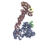
| ||||||||
|---|---|---|---|---|---|---|---|---|---|
| 1 |
| ||||||||
| Unit cell |
|
-Components
| #1: Protein | Mass: 127117.500 Da / Num. of mol.: 1 Source method: isolated from a genetically manipulated source Source: (gene. exp.) Homo sapiens (human) / Gene: DDB1 / Production host:   Spodoptera frugiperda (fall armyworm) / References: UniProt: Q16531 Spodoptera frugiperda (fall armyworm) / References: UniProt: Q16531 | ||
|---|---|---|---|
| #2: Protein | Mass: 23965.039 Da / Num. of mol.: 1 Source method: isolated from a genetically manipulated source Source: (gene. exp.) Simian virus 5 / Genus: Rubulavirus / Gene: P/V / Production host:  | ||
| #3: Protein | Mass: 87814.297 Da / Num. of mol.: 1 Source method: isolated from a genetically manipulated source Source: (gene. exp.) Homo sapiens (human) / Gene: CUL4A / Production host: | ||
| #4: Protein | Mass: 12289.977 Da / Num. of mol.: 1 Source method: isolated from a genetically manipulated source Source: (gene. exp.) Homo sapiens (human) / Gene: RBX1 / Production host: | ||
| #5: Chemical | ChemComp-ZN /   Mass: 65.409 Da / Num. of mol.: 5 / Source method: obtained synthetically / Formula: Zn Mass: 65.409 Da / Num. of mol.: 5 / Source method: obtained synthetically / Formula: ZnHas protein modification | Y | |
-Experimental details
-Experiment
| Experiment | Method: X-RAY DIFFRACTION / Number of used crystals: 1 |
|---|
- Sample preparation
| Crystal | Density Matthews: 3.58 Å3/Da / Density % sol: 65.68 % |
|---|---|
| Crystal grow | Temperature: 277 K / Method: vapor diffusion, hanging drop / pH: 8 Details: 100mM NaHEPES, 7-9% PEG4000, 10% iso-propanol, 5mM DTT, pH 8.0, VAPOR DIFFUSION, HANGING DROP, temperature 277K |
-Data collection
| Diffraction source | Source: SYNCHROTRON / Site: ALS  / Beamline: 8.2.2 / Wavelength: 1 Å / Beamline: 8.2.2 / Wavelength: 1 Å | ||||||||||||||||||||||||||||||||||||||||||||||||||||||||||||||||||
|---|---|---|---|---|---|---|---|---|---|---|---|---|---|---|---|---|---|---|---|---|---|---|---|---|---|---|---|---|---|---|---|---|---|---|---|---|---|---|---|---|---|---|---|---|---|---|---|---|---|---|---|---|---|---|---|---|---|---|---|---|---|---|---|---|---|---|---|
| Radiation | Protocol: SINGLE WAVELENGTH / Monochromatic (M) / Laue (L): M / Scattering type: x-ray | ||||||||||||||||||||||||||||||||||||||||||||||||||||||||||||||||||
| Radiation wavelength | Wavelength: 1 Å / Relative weight: 1 | ||||||||||||||||||||||||||||||||||||||||||||||||||||||||||||||||||
| Reflection | Resolution: 3.1→50 Å / Num. obs: 62466 / % possible obs: 92 % / Redundancy: 5.4 % / Rmerge(I) obs: 0.079 / Χ2: 1.985 / Net I/σ(I): 12.2 | ||||||||||||||||||||||||||||||||||||||||||||||||||||||||||||||||||
| Reflection shell |
|
- Processing
| Software |
| ||||||||||||||||||||
|---|---|---|---|---|---|---|---|---|---|---|---|---|---|---|---|---|---|---|---|---|---|
| Refinement | Method to determine structure: MOLECULAR REPLACEMENT / Resolution: 3.1→50 Å / σ(F): 0 / Stereochemistry target values: Engh & Huber
| ||||||||||||||||||||
| Displacement parameters | Biso mean: 75.92 Å2 | ||||||||||||||||||||
| Refinement step | Cycle: LAST / Resolution: 3.1→50 Å
|
