 Movie
Movie Controller


+ Open data

- Basic information
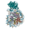
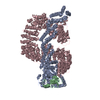
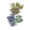
| Entry | Database: PDB / ID: 4a0c | ||||||
|---|---|---|---|---|---|---|---|
| Title | Structure of the CAND1-CUL4B-RBX1 complex | ||||||
 Components Components |
| ||||||
 Keywords Keywords | CELL CYCLE / TRANSCRIPTION / LIGASE / UBIQUITIN / DNA DAMAGE REPAIR | ||||||
| Function / homology |  Function and homology information Function and homology informationProlactin receptor signaling / SCF complex assembly / Recognition of DNA damage by PCNA-containing replication complex / Formation of TC-NER Pre-Incision Complex / DNA Damage Recognition in GG-NER / Dual Incision in GG-NER / Dual incision in TC-NER / Gap-filling DNA repair synthesis and ligation in TC-NER / Formation of Incision Complex in GG-NER / Regulation of BACH1 activity ...Prolactin receptor signaling / SCF complex assembly / Recognition of DNA damage by PCNA-containing replication complex / Formation of TC-NER Pre-Incision Complex / DNA Damage Recognition in GG-NER / Dual Incision in GG-NER / Dual incision in TC-NER / Gap-filling DNA repair synthesis and ligation in TC-NER / Formation of Incision Complex in GG-NER / Regulation of BACH1 activity / Regulation of RAS by GAPs / Degradation of DVL / Ubiquitin-Mediated Degradation of Phosphorylated Cdc25A / Regulation of RUNX2 expression and activity / Degradation of GLI1 by the proteasome / Hedgehog 'on' state / FBXL7 down-regulates AURKA during mitotic entry and in early mitosis / Orc1 removal from chromatin / GSK3B and BTRC:CUL1-mediated-degradation of NFE2L2 / eukaryotic initiation factor 4E binding / Oxygen-dependent proline hydroxylation of Hypoxia-inducible Factor Alpha / negative regulation of catalytic activity / Degradation of beta-catenin by the destruction complex / base-excision repair, AP site formation via deaminated base removal / miRNA-mediated gene silencing by mRNA destabilization / anaphase-promoting complex / Interleukin-1 signaling / GLI3 is processed to GLI3R by the proteasome / Neddylation / cullin-RING-type E3 NEDD8 transferase / astrocyte differentiation / KEAP1-NFE2L2 pathway / cullin-RING ubiquitin ligase complex / Cul7-RING ubiquitin ligase complex / Antigen processing: Ubiquitination & Proteasome degradation / positive regulation of protein autoubiquitination / RNA polymerase II transcription initiation surveillance / positive regulation of epithelial cell apoptotic process / protein neddylation / NEDD8 ligase activity / lysosome organization / negative regulation of response to oxidative stress / UV-damage excision repair / VCB complex / Cul5-RING ubiquitin ligase complex / ubiquitin-ubiquitin ligase activity / ubiquitin-dependent protein catabolic process via the C-end degron rule pathway / SCF ubiquitin ligase complex / Cul2-RING ubiquitin ligase complex / type I interferon-mediated signaling pathway / Cul3-RING ubiquitin ligase complex / negative regulation of type I interferon production / SCF-dependent proteasomal ubiquitin-dependent protein catabolic process / p38MAPK cascade / Cul4A-RING E3 ubiquitin ligase complex / Cul4-RING E3 ubiquitin ligase complex / Cul4B-RING E3 ubiquitin ligase complex / negative regulation of signal transduction by p53 class mediator / TORC1 signaling / epithelial to mesenchymal transition / positive regulation of RNA polymerase II transcription preinitiation complex assembly / cullin family protein binding / protein monoubiquitination / ubiquitin ligase complex / positive regulation of G1/S transition of mitotic cell cycle / negative regulation of ubiquitin-dependent protein catabolic process / autophagosome assembly / mitophagy / site of DNA damage / positive regulation of type I interferon production / signal transduction in response to DNA damage / negative regulation of TORC1 signaling / protein K48-linked ubiquitination / negative regulation of insulin receptor signaling pathway / positive regulation of TORC1 signaling / negative regulation of proteasomal ubiquitin-dependent protein catabolic process / regulation of mitotic cell cycle / transcription-coupled nucleotide-excision repair / positive regulation of autophagy / cellular response to nutrient levels / TBP-class protein binding / cellular response to amino acid starvation / protein catabolic process / negative regulation of autophagy / negative regulation of canonical NF-kappaB signal transduction / T cell activation / DNA damage response, signal transduction by p53 class mediator / positive regulation of translation / cellular response to amino acid stimulus / proteasomal protein catabolic process / G1/S transition of mitotic cell cycle / transcription elongation by RNA polymerase II / Iron uptake and transport / Recognition of DNA damage by PCNA-containing replication complex / RING-type E3 ubiquitin transferase / DNA Damage Recognition in GG-NER / Dual Incision in GG-NER / Transcription-Coupled Nucleotide Excision Repair (TC-NER) / Formation of TC-NER Pre-Incision Complex / cellular response to insulin stimulus Similarity search - Function | ||||||
| Biological species |  HOMO SAPIENS (human) HOMO SAPIENS (human) | ||||||
| Method |  X-RAY DIFFRACTION / SYNCHROTRON / MOLECULAR REPLACEMENT / Resolution: 3.8 Å X-RAY DIFFRACTION / SYNCHROTRON / MOLECULAR REPLACEMENT / Resolution: 3.8 Å | ||||||
 Authors Authors | Scrima, A. / Fischer, E.S. / Faty, M. / Gut, H. / Thoma, N.H. | ||||||
 Citation Citation | Journal: Cell(Cambridge,Mass.) / Year: 2011 Title: The Molecular Basis of Crl4(Ddb2/Csa) Ubiquitin Ligase Architecture, Targeting, and Activation Authors: Scrima, A. / Fischer, E.S. / Iwai, S. / Gut, H. / Thoma, N.H. | ||||||
| History |
|
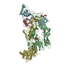
- Structure visualization
| Structure viewer | Molecule: MolmilJmol/JSmol |
|---|
- Downloads & links
-Download
| PDBx/mmCIF format | 4a0c.cif.gz | 755.4 KB | Display | PDBx/mmCIF format |
|---|---|---|---|---|
| PDB format | pdb4a0c.ent.gz | 604 KB | Display | PDB format |
| PDBx/mmJSON format | 4a0c.json.gz | Tree view | PDBx/mmJSON format | |
| Others |  Other downloads Other downloads |
-Validation report
| Arichive directory | https://data.pdbj.org/pub/pdb/validation_reports/a0/4a0cftp://data.pdbj.org/pub/pdb/validation_reports/a0/4a0c | HTTPS FTP |
|---|
-Related structure data
| Related structure data | 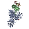 4a08C 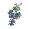 4a09C 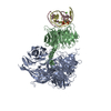 4a0aC 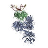 4a0bC 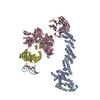 4a0kC 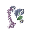 4a0lC  4a11C  1u6gS 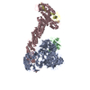 2hyeS S: Starting model for refinement C: citing same article ( |
|---|---|
| Similar structure data |


-Links
 PDBj
PDBj


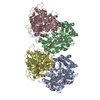
- Assembly
| Deposited unit | 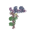
| ||||||||
|---|---|---|---|---|---|---|---|---|---|
| 1 | 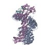
| ||||||||
| 2 | 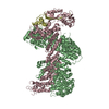
| ||||||||
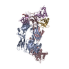
| Unit cell |
|
-Components
| #1: Protein | Mass: 139226.281 Da / Num. of mol.: 2 Source method: isolated from a genetically manipulated source Source: (gene. exp.) HOMO SAPIENS (human) / Plasmid: PFASTBAC DERIVED / Cell line (production host): High Five / Production host:  TRICHOPLUSIA NI (cabbage looper) / References: UniProt: Q86VP6 TRICHOPLUSIA NI (cabbage looper) / References: UniProt: Q86VP6#2: Protein | Mass: 86961.336 Da / Num. of mol.: 2 Source method: isolated from a genetically manipulated source Source: (gene. exp.) HOMO SAPIENS (human) / Plasmid: PFASTBAC DERIVED / Cell line (production host): High Five / Production host: TRICHOPLUSIA NI (cabbage looper) / References: UniProt: Q13620#3: Protein | Mass: 11330.942 Da / Num. of mol.: 2 Source method: isolated from a genetically manipulated source Source: (gene. exp.) TRICHOPLUSIA NI (cabbage looper)References: UniProt: P62878, Ligases; Forming carbon-nitrogen bonds; Acid-amino-acid ligases (peptide synthases) #4: Chemical | ChemComp-ZN /   Mass: 65.409 Da / Num. of mol.: 6 / Source method: obtained synthetically / Formula: Zn Mass: 65.409 Da / Num. of mol.: 6 / Source method: obtained synthetically / Formula: ZnSequence details | CHAINS A AND B CONTAINS NATURAL VARIANT A952V | |
|---|
-Experimental details
-Experiment
| Experiment | Method: X-RAY DIFFRACTION / Number of used crystals: 1 |
|---|
- Sample preparation
| Crystal | Density Matthews: 3.52 Å3/Da / Density % sol: 65.08 % Description: CAND1 USED AS MODEL FROM 1U6G, CUL4B MODEL GENERATED USING MODELLER BASED ON CUL4A FROM 2HYE |
|---|---|
| Crystal grow | pH: 6.3 / Details: 100 MM MES PH 6.3, 30% PEG 200, 2% PEG 8000. |
-Data collection
| Diffraction | Mean temperature: 100 K |
|---|---|
| Diffraction source | Source: SYNCHROTRON / Site: SLS  / Beamline: X10SA / Wavelength: 1 / Beamline: X10SA / Wavelength: 1 |
| Detector | Type: DECTRIS PILATUS 6M / Detector: PIXEL / Date: Jul 30, 2010 |
| Radiation | Protocol: SINGLE WAVELENGTH / Monochromatic (M) / Laue (L): M / Scattering type: x-ray |
| Radiation wavelength | Wavelength: 1 Å / Relative weight: 1 |
| Reflection | Resolution: 3.8→50 Å / Num. obs: 59850 / % possible obs: 99.7 % / Observed criterion σ(I): 2 / Redundancy: 3.8 % / Rmerge(I) obs: 0.09 / Net I/σ(I): 10.2 |
| Reflection shell | Resolution: 3.8→3.9 Å / Redundancy: 3.8 % / Rmerge(I) obs: 0.51 / Mean I/σ(I) obs: 2.8 / % possible all: 99.8 |
- Processing
| Software |
| ||||||||||||||||||||||||||||||||||||||||||||||||||||||||||||||||||||||||||||||||||||||||||||||||||||||||||||||||||||||||||||||||||||||||||||||||||||||||||||||||||||||||||||||||||||||
|---|---|---|---|---|---|---|---|---|---|---|---|---|---|---|---|---|---|---|---|---|---|---|---|---|---|---|---|---|---|---|---|---|---|---|---|---|---|---|---|---|---|---|---|---|---|---|---|---|---|---|---|---|---|---|---|---|---|---|---|---|---|---|---|---|---|---|---|---|---|---|---|---|---|---|---|---|---|---|---|---|---|---|---|---|---|---|---|---|---|---|---|---|---|---|---|---|---|---|---|---|---|---|---|---|---|---|---|---|---|---|---|---|---|---|---|---|---|---|---|---|---|---|---|---|---|---|---|---|---|---|---|---|---|---|---|---|---|---|---|---|---|---|---|---|---|---|---|---|---|---|---|---|---|---|---|---|---|---|---|---|---|---|---|---|---|---|---|---|---|---|---|---|---|---|---|---|---|---|---|---|---|---|---|
| Refinement | Method to determine structure: MOLECULAR REPLACEMENT Starting model: PDB ENTRIES 1U6G, 2HYE Resolution: 3.8→47.76 Å / Cor.coef. Fo:Fc: 0.929 / Cor.coef. Fo:Fc free: 0.882 / SU B: 56.264 / SU ML: 0.797 / Cross valid method: THROUGHOUT / ESU R Free: 0.906 / Stereochemistry target values: MAXIMUM LIKELIHOOD Details: HYDROGENS HAVE BEEN ADDED IN THE RIDING POSITIONS.U VALUES REFINED INDIVIDUALLY.
| ||||||||||||||||||||||||||||||||||||||||||||||||||||||||||||||||||||||||||||||||||||||||||||||||||||||||||||||||||||||||||||||||||||||||||||||||||||||||||||||||||||||||||||||||||||||
| Solvent computation | Ion probe radii: 0.8 Å / Shrinkage radii: 0.8 Å / VDW probe radii: 1.4 Å / Solvent model: MASK | ||||||||||||||||||||||||||||||||||||||||||||||||||||||||||||||||||||||||||||||||||||||||||||||||||||||||||||||||||||||||||||||||||||||||||||||||||||||||||||||||||||||||||||||||||||||
| Displacement parameters | Biso mean: 128.578 Å2
| ||||||||||||||||||||||||||||||||||||||||||||||||||||||||||||||||||||||||||||||||||||||||||||||||||||||||||||||||||||||||||||||||||||||||||||||||||||||||||||||||||||||||||||||||||||||
| Refinement step | Cycle: LAST / Resolution: 3.8→47.76 Å
| ||||||||||||||||||||||||||||||||||||||||||||||||||||||||||||||||||||||||||||||||||||||||||||||||||||||||||||||||||||||||||||||||||||||||||||||||||||||||||||||||||||||||||||||||||||||
| Refine LS restraints |
|