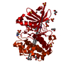
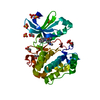
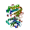
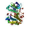
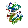
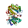
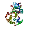
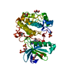
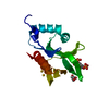
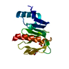
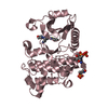
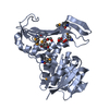
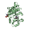
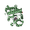
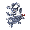
[English] 日本語
Yorodumi
- PDB-4a07: Human PDK1 Kinase Domain in Complex with Allosteric Activator PS1... -

+
Open data


ID or keywords:

Loading...

-
Basic information

Entry
Database: PDB / ID: 4a07
TitleHuman PDK1 Kinase Domain in Complex with Allosteric Activator PS171 Bound to the PIF-Pocket
Components3-PHOSPHOINOSITIDE-DEPENDENT PROTEIN KINASE 1
KeywordsTRANSFERASE / ALLOSTERIC SITE
Function / homology
Function and homology information


3-phosphoinositide-dependent protein kinase activity / Activation of AKT2 / regulation of mast cell degranulation / negative regulation of toll-like receptor signaling pathway / type B pancreatic cell development / positive regulation of phospholipase activity / RSK activation / hyperosmotic response / regulation of canonical NF-kappaB signal transduction / positive regulation of vascular endothelial cell proliferation ...3-phosphoinositide-dependent protein kinase activity / Activation of AKT2 / regulation of mast cell degranulation / negative regulation of toll-like receptor signaling pathway / type B pancreatic cell development / positive regulation of phospholipase activity / RSK activation / hyperosmotic response / regulation of canonical NF-kappaB signal transduction / positive regulation of vascular endothelial cell proliferation / negative regulation of cardiac muscle cell apoptotic process / phospholipase activator activity / positive regulation of sprouting angiogenesis / Constitutive Signaling by AKT1 E17K in Cancer / CD28 dependent PI3K/Akt signaling / phospholipase binding / positive regulation of blood vessel endothelial cell migration / Role of LAT2/NTAL/LAB on calcium mobilization / Estrogen-stimulated signaling through PRKCZ / SARS-CoV-2 targets host intracellular signalling and regulatory pathways / negative regulation of endothelial cell apoptotic process / SARS-CoV-1 targets host intracellular signalling and regulatory pathways / extrinsic apoptotic signaling pathway / RHO GTPases activate PKNs / cellular response to epidermal growth factor stimulus / GPVI-mediated activation cascade / T cell costimulation / activation of protein kinase B activity / Integrin signaling / insulin-like growth factor receptor signaling pathway / positive regulation of release of sequestered calcium ion into cytosol / VEGFR2 mediated vascular permeability / VEGFR2 mediated cell proliferation / cell projection / calcium-mediated signaling / positive regulation of protein localization to plasma membrane / peptidyl-threonine phosphorylation / negative regulation of transforming growth factor beta receptor signaling pathway / negative regulation of protein kinase activity / epidermal growth factor receptor signaling pathway / CLEC7A (Dectin-1) signaling / FCERI mediated NF-kB activation / G beta:gamma signalling through PI3Kgamma / cellular response to insulin stimulus / positive regulation of angiogenesis / Regulation of TP53 Degradation / cell migration / Downstream TCR signaling / insulin receptor signaling pathway / PIP3 activates AKT signaling / cytoplasmic vesicle / actin cytoskeleton organization / protein autophosphorylation / postsynaptic density / positive regulation of phosphatidylinositol 3-kinase/protein kinase B signal transduction / non-specific serine/threonine protein kinase / intracellular signal transduction / protein phosphorylation / protein serine kinase activity / focal adhesion / protein serine/threonine kinase activity / ATP binding / nucleus / plasma membrane / cytoplasm / cytosol
Similarity search - Function
PDK1-type, PH domain / PDPK1 family / PH domain / PH-like domain superfamily / Transferase(Phosphotransferase) domain 1 / Transferase(Phosphotransferase); domain 1 / Phosphorylase Kinase; domain 1 / Phosphorylase Kinase; domain 1 / Serine/threonine-protein kinase, active site / Serine/Threonine protein kinases active-site signature. ...PDK1-type, PH domain / PDPK1 family / PH domain / PH-like domain superfamily / Transferase(Phosphotransferase) domain 1 / Transferase(Phosphotransferase); domain 1 / Phosphorylase Kinase; domain 1 / Phosphorylase Kinase; domain 1 / Serine/threonine-protein kinase, active site / Serine/Threonine protein kinases active-site signature. / Protein kinase domain / Serine/Threonine protein kinases, catalytic domain / Protein kinase, ATP binding site / Protein kinases ATP-binding region signature. / Protein kinase domain profile. / Protein kinase domain / Protein kinase-like domain superfamily / 2-Layer Sandwich / Orthogonal Bundle / Mainly Alpha / Alpha Beta
Similarity search - Domain/homology
ADENOSINE-5'-TRIPHOSPHATE / Chem-AZ7 / 3-phosphoinositide-dependent protein kinase 1
Similarity search - Component
Biological speciesHOMO SAPIENS (human)
MethodX-RAY DIFFRACTION / SYNCHROTRON / MOLECULAR REPLACEMENT / Resolution: 1.85 Å
AuthorsSchulze, J.O. / Lopez-Garcia, L.A. / Froehner, W. / Zhang, H. / Navratil, J. / Hindie, V. / Zeuzem, S. / Alzari, P.M. / Neimanis, S. / Engel, M. / Biondi, R.M.
CitationJournal: Chem.Biol. / Year: 2011
Title: Allosteric Regulation of Protein Kinase Pkczeta by the N-Terminal C1 Domain and Small Compounds to the Pif-Pocket.
Authors: Lopez-Garcia, L.A. / Schulze, J.O. / Frohner, W. / Zhang, H. / Suss, E. / Weber, N. / Navratil, J. / Amon, S. / Hindie, V. / Zeuzem, S. / Jorgensen, T.J. / Alzari, P.M. / Neimanis, S. / Engel, M. / Biondi, R.M.
History
DepositionSep 8, 2011Deposition site: PDBE / Processing site: PDBE
Revision 1.0Dec 7, 2011Provider: repository / Type: Initial release
Revision 1.1Dec 21, 2011Group: Other
Revision 1.2Dec 20, 2023Group: Data collection / Database references ...Data collection / Database references / Derived calculations / Other / Refinement description
Category: chem_comp_atom / chem_comp_bond ...chem_comp_atom / chem_comp_bond / database_2 / pdbx_database_status / pdbx_initial_refinement_model / pdbx_struct_conn_angle / struct_conn / struct_site
Item: _database_2.pdbx_DOI / _database_2.pdbx_database_accession ..._database_2.pdbx_DOI / _database_2.pdbx_database_accession / _pdbx_database_status.status_code_sf / _pdbx_struct_conn_angle.ptnr1_auth_comp_id / _pdbx_struct_conn_angle.ptnr1_auth_seq_id / _pdbx_struct_conn_angle.ptnr1_label_asym_id / _pdbx_struct_conn_angle.ptnr1_label_atom_id / _pdbx_struct_conn_angle.ptnr1_label_comp_id / _pdbx_struct_conn_angle.ptnr1_label_seq_id / _pdbx_struct_conn_angle.ptnr3_auth_comp_id / _pdbx_struct_conn_angle.ptnr3_auth_seq_id / _pdbx_struct_conn_angle.ptnr3_label_asym_id / _pdbx_struct_conn_angle.ptnr3_label_atom_id / _pdbx_struct_conn_angle.ptnr3_label_comp_id / _pdbx_struct_conn_angle.ptnr3_label_seq_id / _pdbx_struct_conn_angle.value / _struct_conn.pdbx_dist_value / _struct_conn.pdbx_leaving_atom_flag / _struct_conn.ptnr1_auth_comp_id / _struct_conn.ptnr1_auth_seq_id / _struct_conn.ptnr1_label_asym_id / _struct_conn.ptnr1_label_atom_id / _struct_conn.ptnr1_label_comp_id / _struct_conn.ptnr1_label_seq_id / _struct_conn.ptnr2_auth_comp_id / _struct_conn.ptnr2_auth_seq_id / _struct_conn.ptnr2_label_asym_id / _struct_conn.ptnr2_label_atom_id / _struct_conn.ptnr2_label_comp_id / _struct_conn.ptnr2_label_seq_id / _struct_site.pdbx_auth_asym_id / _struct_site.pdbx_auth_comp_id / _struct_site.pdbx_auth_seq_id
Remark 650 HELIX DETERMINATION METHOD: AUTHOR PROVIDED.
Remark 700 SHEET DETERMINATION METHOD: AUTHOR PROVIDED.

-
Structure visualization

Structure viewerMolecule:
MolmilJmol/JSmol

Downloads & links

-
Assembly

Deposited unit
A: 3-PHOSPHOINOSITIDE-DEPENDENT PROTEIN KINASE 1
hetero molecules


Theoretical massNumber of molelcules
Total (without water)36,90110
Polymers35,4251
Non-polymers1,4769
Water3,765209
1


  • Idetical with deposited unit
  • defined by author&software
TypeNameSymmetry operationNumber
identity operation1_555x,y,z1
MethodPISA
Unit cell
Length a, b, c (Å)148.210, 44.030, 47.670
Angle α, β, γ (deg.)90.00, 101.23, 90.00
Int Tables number5
Space group name H-MC121

-
Components

-
Protein , 1 types, 1 molecules A

#1: Protein 3-PHOSPHOINOSITIDE-DEPENDENT PROTEIN KINASE 1 / HPDK1


Mass: 35424.629 Da / Num. of mol.: 1 / Fragment: CATALYTIC DOMAIN, RESIDUES 50-359 / Mutation: YES
Source method: isolated from a genetically manipulated source
Source: (gene. exp.) HOMO SAPIENS (human) / Cell line (production host): SF9 / Production host: 
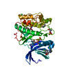
SPODOPTERA FRUGIPERDA (fall armyworm)
References: UniProt: O15530, non-specific serine/threonine protein kinase

-
Non-polymers , 5 types, 218 molecules

#2: Chemical ChemComp-ATP / ADENOSINE-5'-TRIPHOSPHATE


Mass: 507.181 Da / Num. of mol.: 1 / Source method: obtained synthetically / Formula: C10H16N5O13P3 / Comment: ATP, energy-carrying molecule*YM
#3: Chemical ChemComp-AZ7 / (3S)-3-(4-CHLOROPHENYL)-4-(5,7-DICHLORO-1H-BENZIMIDAZOL-2-YL)BUTANOIC ACID


Mass: 383.656 Da / Num. of mol.: 2 / Source method: obtained synthetically / Formula: C17H13Cl3N2O2
#4: Chemical ChemComp-MG / MAGNESIUM ION


Mass: 24.305 Da / Num. of mol.: 1 / Source method: obtained synthetically / Formula: Mg
#5: Chemical
ChemComp-CL / CHLORIDE ION


Mass: 35.453 Da / Num. of mol.: 5 / Source method: obtained synthetically / Formula: Cl
#6: Water ChemComp-HOH / water


Mass: 18.015 Da / Num. of mol.: 209 / Source method: isolated from a natural source / Formula: H2O

-
Details

Compound detailsENGINEERED RESIDUE IN CHAIN A, GLN 288 TO ALA ENGINEERED RESIDUE IN CHAIN A, TYR 292 TO GLY
Nonpolymer detailsPS171 (AZ7): ALLOSTERIC ACTIVATOR PS171

-
Experimental details

-
Experiment

ExperimentMethod: X-RAY DIFFRACTION / Number of used crystals: 1

-
Sample preparation

CrystalDensity Matthews: 2.3 Å3/Da / Density % sol: 47 % / Description: NONE
Crystal growpH: 7.5 / Details: 1.2 M NA CITRATE 0.1 M HEPES PH 7.5 10 M DTT.

-
Data collection

DiffractionMean temperature: 100 K
Diffraction sourceSource: SYNCHROTRON / Site: SLS / Beamline: X06DA / Wavelength: 0.9999
DetectorType: RAYONIX / Detector: CCD / Date: Dec 4, 2009 / Details: MIRRORS
RadiationProtocol: SINGLE WAVELENGTH / Monochromatic (M) / Laue (L): M / Scattering type: x-ray
Radiation wavelengthWavelength: 0.9999 Å / Relative weight: 1
ReflectionResolution: 1.85→47 Å / Num. obs: 25852 / % possible obs: 99.3 % / Observed criterion σ(I): 2.8 / Redundancy: 4.6 % / Biso Wilson estimate: 16.59 Å2 / Rmerge(I) obs: 0.1 / Net I/σ(I): 15.5
Reflection shellResolution: 1.85→1.95 Å / Redundancy: 4.6 % / Rmerge(I) obs: 0.72 / Mean I/σ(I) obs: 2.8 / % possible all: 98.9

-
Processing

Software
NameVersionClassification
PHENIX(PHENIX.REFINE)refinement
XDSdata reduction
XSCALEdata scaling
PHASERphasing
RefinementMethod to determine structure: MOLECULAR REPLACEMENT
Starting model: PDB ENTRY 3HRC
Resolution: 1.85→36.242 Å / SU ML: 0.19 / σ(F): 2 / Phase error: 18.42 / Stereochemistry target values: ML
RfactorNum. reflection% reflection
Rfree0.1997 1292 5 %
Rwork0.1588 --
obs0.1609 25850 99.37 %
Solvent computationShrinkage radii: 0.89 Å / VDW probe radii: 1 Å / Solvent model: FLAT BULK SOLVENT MODEL / Bsol: 40.291 Å2 / ksol: 0.426 e/Å3
Displacement parameters
Baniso -1Baniso -2Baniso -3
1--0.5851 Å20 Å2-1.4489 Å2
2--0.2149 Å20 Å2
3---0.3702 Å2
Refinement stepCycle: LAST / Resolution: 1.85→36.242 Å
ProteinNucleic acidLigandSolventTotal
Num. atoms2240 0 85 209 2534
Refine LS restraints
Refine-IDTypeDev idealNumber
X-RAY DIFFRACTIONf_bond_d0.0082417
X-RAY DIFFRACTIONf_angle_d1.093308
X-RAY DIFFRACTIONf_dihedral_angle_d13.644884
X-RAY DIFFRACTIONf_chiral_restr0.074356
X-RAY DIFFRACTIONf_plane_restr0.005409
LS refinement shell
Resolution (Å)Rfactor RfreeNum. reflection RfreeRfactor RworkNum. reflection RworkRefine-ID% reflection obs (%)
1.8499-1.9240.27681410.22612670X-RAY DIFFRACTION99
1.924-2.01150.2161410.18332692X-RAY DIFFRACTION99
2.0115-2.11760.23331420.15652692X-RAY DIFFRACTION99
2.1176-2.25020.17991430.14672717X-RAY DIFFRACTION99
2.2502-2.42390.18451430.14322722X-RAY DIFFRACTION99
2.4239-2.66780.20381440.15442740X-RAY DIFFRACTION100
2.6678-3.05360.21931440.15872723X-RAY DIFFRACTION100
3.0536-3.84660.17761450.15342762X-RAY DIFFRACTION100
3.8466-36.24860.18831490.15562840X-RAY DIFFRACTION100
Refinement TLS params.

Method: refined / Refine-ID: X-RAY DIFFRACTION

IDL112)L122)L132)L222)L232)L332)S11 (Å °)S12 (Å °)S13 (Å °)S21 (Å °)S22 (Å °)S23 (Å °)S31 (Å °)S32 (Å °)S33 (Å °)T112)T122)T132)T222)T232)T332)Origin x (Å)Origin y (Å)Origin z (Å)
12.29462.17820.39193.07990.24111.25320.12670.16560.2069-0.1657-0.13220.30110.0217-0.2107-0.01080.10350.0301-0.01860.210.03830.104-8.915524.5124-16.1908
20.1087-0.0676-0.24820.26670.64241.74990.00380.1490.0186-0.0325-0.0548-0.0423-0.11620.16320.06490.06790.0353-0.00780.20260.0330.0451-7.093325.6663-13.9567
32.7577-0.82620.31220.70960.49081.20120.46340.2953-0.072-0.1464-0.31110.1598-0.1858-0.3181-0.10380.18360.07080.01530.23650.01690.1795-16.300934.6489-4.4123
40.2466-0.03760.23950.8801-0.14311.8736-0.02940.03820.1257-0.0345-0.0186-0.0981-0.3676-0.04020.03180.1305-0.01760.00660.12740.02460.0846-7.356826.4935-3.8426
50.1774-0.2215-0.11411.6271-0.27070.31560.10540.07840.1141-0.1855-0.2508-0.0112-0.02510.080.10820.12430.0286-0.00290.15020.02020.0818-10.186324.5599-11.3339
60.91270.27920.10130.62440.36430.7323-0.05360.0098-0.11060.03860.0786-0.06610.04740.0707-0.04550.06870.0022-0.00460.0762-0.00880.086-19.903314.88095.681
71.0562-0.7376-0.36021.4892-0.09780.867-0.01610.08270.1239-0.04450.0051-0.24-0.16530.07020.02570.089-0.0239-0.0120.12330.00010.0835-17.003425.13924.0766
80.84480.0684-0.09520.2345-0.03410.6407-0.0518-0.01930.1327-0.02040.01490.1823-0.13570.04140.04080.09090.0053-0.02250.06450.00860.1002-30.792122.60656.5538
90.8385-0.180.25141.0455-0.14790.8777-0.0333-0.1049-0.12360.09760.010.24320.106-0.0920.01560.0895-0.00880.02670.07380.00530.1253-35.177915.5211.3834
100.59060.3334-0.12861.7423-0.64380.27230.1264-0.11550.06880.4545-0.1383-0.046-0.20710.13770.03720.18170.0111-0.05280.1537-0.00220.046-21.516222.232621.2906
110.8660.2832-0.5420.8243-0.13122.1632-0.3661-0.0517-0.42560.13170.0964-0.33490.44860.28680.14760.17380.0407-0.00080.16750.01750.1966-13.12278.963314.279
Refinement TLS group
IDRefine-IDRefine TLS-IDSelection details
1X-RAY DIFFRACTION1(CHAIN A AND RESID 75:99)
2X-RAY DIFFRACTION2(CHAIN A AND RESID 100:117)
3X-RAY DIFFRACTION3(CHAIN A AND RESID 118:138)
4X-RAY DIFFRACTION4(CHAIN A AND RESID 139:151)
5X-RAY DIFFRACTION5(CHAIN A AND RESID 152:165)
6X-RAY DIFFRACTION6(CHAIN A AND RESID 166:208)
7X-RAY DIFFRACTION7(CHAIN A AND RESID 209:238)
8X-RAY DIFFRACTION8(CHAIN A AND RESID 239:283)
9X-RAY DIFFRACTION9(CHAIN A AND RESID 284:325)
10X-RAY DIFFRACTION10(CHAIN A AND RESID 326:338)
11X-RAY DIFFRACTION11(CHAIN A AND RESID 339:357)

+
About Yorodumi

-
News

-
Feb 9, 2022. New format data for meta-information of EMDB entries

New format data for meta-information of EMDB entries

  • Version 3 of the EMDB header file is now the official format.
  • The previous official version 1.9 will be removed from the archive.

Related info.:EMDB header

External links:wwPDB to switch to version 3 of the EMDB data model

-
Aug 12, 2020. Covid-19 info

Covid-19 info

URL: https://pdbjlvh1.pdbj.org/emnavi/covid19.php

New page: Covid-19 featured information page in EM Navigator.

Related info.:Covid-19 info / Mar 5, 2020. Novel coronavirus structure data

+
Mar 5, 2020. Novel coronavirus structure data

Novel coronavirus structure data

Related info.:Yorodumi Speices / Aug 12, 2020. Covid-19 info

External links:COVID-19 featured content - PDBj / Molecule of the Month (242):Coronavirus Proteases

+
Jan 31, 2019. EMDB accession codes are about to change! (news from PDBe EMDB page)

EMDB accession codes are about to change! (news from PDBe EMDB page)

  • The allocation of 4 digits for EMDB accession codes will soon come to an end. Whilst these codes will remain in use, new EMDB accession codes will include an additional digit and will expand incrementally as the available range of codes is exhausted. The current 4-digit format prefixed with “EMD-” (i.e. EMD-XXXX) will advance to a 5-digit format (i.e. EMD-XXXXX), and so on. It is currently estimated that the 4-digit codes will be depleted around Spring 2019, at which point the 5-digit format will come into force.
  • The EM Navigator/Yorodumi systems omit the EMD- prefix.

Related info.:Q: What is EMD? / ID/Accession-code notation in Yorodumi/EM Navigator

External links:EMDB Accession Codes are Changing Soon! / Contact to PDBj

+
Jul 12, 2017. Major update of PDB

Major update of PDB

  • wwPDB released updated PDB data conforming to the new PDBx/mmCIF dictionary.
  • This is a major update changing the version number from 4 to 5, and with Remediation, in which all the entries are updated.
  • In this update, many items about electron microscopy experimental information are reorganized (e.g. em_software).
  • Now, EM Navigator and Yorodumi are based on the updated data.

External links:wwPDB Remediation / Enriched Model Files Conforming to OneDep Data Standards Now Available in the PDB FTP Archive

-
Yorodumi

Thousand views of thousand structures

  • Yorodumi is a browser for structure data from EMDB, PDB, SASBDB, etc.
  • This page is also the successor to EM Navigator detail page, and also detail information page/front-end page for Omokage search.
  • The word "yorodu" (or yorozu) is an old Japanese word meaning "ten thousand". "mi" (miru) is to see.

Related info.:EMDB / PDB / SASBDB / Comparison of 3 databanks / Yorodumi Search / Aug 31, 2016. New EM Navigator & Yorodumi / Yorodumi Papers / Jmol/JSmol / Function and homology information / Changes in new EM Navigator and Yorodumi

Read more