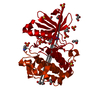
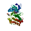
Entry Database : PDB / ID : 3hrcTitle Crystal structure of a mutant of human PDK1 Kinase domain in complex with ATP 3-phosphoinositide-dependent protein kinase 1 Keywords / / / / / / / / / / / / Function / homology Function Domain/homology Component
/ / / / / / / / / / / / / / / / / / / / / / / / / / / / / / / / / / / / / / / / / / / / / / / / / / / / / / / / / / / / / / / / / / / / / / / / / / / / / / / / / / / / / / / / / / / / / / / / / / / / / / / / / Biological species Homo sapiens (human)Method / / / / Resolution : 1.91 Å Authors Hindie, V. / Alzari, P.M. / Biondi, R.M. Journal : Nat.Chem.Biol. / Year : 2009Title : Structure and allosteric effects of low-molecular-weight activators on the protein kinase PDK1.Authors : Hindie, V. / Stroba, A. / Zhang, H. / Lopez-Garcia, L.A. / Idrissova, L. / Zeuzem, S. / Hirschberg, D. / Schaeffer, F. / Jrgensen, T.J. / Engel, M. / Alzari, P.M. / Biondi, R.M. History Deposition Jun 9, 2009 Deposition site / Processing site Revision 1.0 Sep 15, 2009 Provider / Type Revision 1.1 Jul 13, 2011 Group / Version format complianceRevision 1.2 Nov 13, 2013 Group Revision 1.3 Nov 10, 2021 Group / Derived calculationsCategory database_2 / struct_conn ... database_2 / struct_conn / struct_ref_seq_dif / struct_site Item _database_2.pdbx_DOI / _database_2.pdbx_database_accession ... _database_2.pdbx_DOI / _database_2.pdbx_database_accession / _struct_conn.pdbx_leaving_atom_flag / _struct_ref_seq_dif.details / _struct_site.pdbx_auth_asym_id / _struct_site.pdbx_auth_comp_id / _struct_site.pdbx_auth_seq_id Revision 1.4 Nov 1, 2023 Group / Refinement descriptionCategory / chem_comp_bond / pdbx_initial_refinement_modelRevision 1.5 Nov 20, 2024 Group / Category / pdbx_modification_feature / Item
Show all Show less
 Movie
Movie Controller

 Yorodumi
 Open data
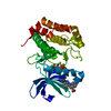
 Basic information
 Components
 Keywords
 Function and homology information
 Homo sapiens (human)
 X-RAY DIFFRACTION /
  Authors
 Citation
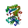
 Structure visualization
 Downloads & links
 Other downloads


 PDBj
PDBj


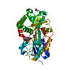
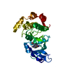
 Assembly
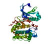

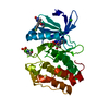
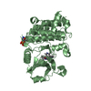
 Spodoptera frugiperda (fall armyworm)
 Mass: 507.181 Da / Num. of mol.: 1 / Source method: obtained synthetically / Formula: C10H16N5O13P3 / Comment: ATP, energy-carrying molecule*YM
 Mass: 18.015 Da / Num. of mol.: 178 / Source method: isolated from a natural source / Formula: H2O
 Sample preparation
 / Beamline: ID14-1 / Wavelength: 0.934 Å
 Processing