 Movie
Movie Controller

[English] 日本語
 Yorodumi
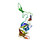
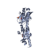
- PDB-3zhg: Crystallographic structure of the native mouse SIGN-R1 CRD domain -
+ Open data

- Basic information
| Entry | Database: PDB / ID: 3zhg | ||||||
|---|---|---|---|---|---|---|---|
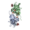
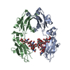
| Title | Crystallographic structure of the native mouse SIGN-R1 CRD domain | ||||||
 Components Components | (CD209 ANTIGEN-LIKE PROTEIN B) x 2 | ||||||
 Keywords Keywords | IMMUNE SYSTEM / C-LECTIN CRD / CAPSULAR POLYSACCHARIDE. | ||||||
| Function / homology |  Function and homology information Function and homology informationdetection of yeast / (1->3)-beta-D-glucan binding / phagocytosis, recognition / pattern recognition receptor activity / D-mannose binding / polysaccharide binding / detection of bacterium / positive regulation of phagocytosis / endocytosis / positive regulation of tumor necrosis factor production ...detection of yeast / (1->3)-beta-D-glucan binding / phagocytosis, recognition / pattern recognition receptor activity / D-mannose binding / polysaccharide binding / detection of bacterium / positive regulation of phagocytosis / endocytosis / positive regulation of tumor necrosis factor production / calcium-dependent protein binding / virus receptor activity / immune response / external side of plasma membrane / membrane / metal ion binding / plasma membrane Similarity search - Function | ||||||
| Biological species |  | ||||||
| Method |  X-RAY DIFFRACTION / SYNCHROTRON / MOLECULAR REPLACEMENT / Resolution: 1.87 Å X-RAY DIFFRACTION / SYNCHROTRON / MOLECULAR REPLACEMENT / Resolution: 1.87 Å | ||||||
 Authors Authors | Silva-Martin, N. / Bartual, S.G. / Hermoso, J.A. | ||||||
 Citation Citation | Journal: Structure / Year: 2014 Title: Structural Basis for Selective Recognition of Endogenous and Microbial Polysaccharides by Macrophage Receptor Sign-R1. Authors: Silva-Martin, N. / Bartual, S.G. / Ramirez-Aportela, E. / Chacon, P. / Park, C.G. / Hermoso, J.A. | ||||||
| History |
|
- Structure visualization
| Structure viewer | Molecule: MolmilJmol/JSmol |
|---|
- Downloads & links
-Download
| PDBx/mmCIF format | 3zhg.cif.gz | 134.2 KB | Display | PDBx/mmCIF format |
|---|---|---|---|---|
| PDB format | pdb3zhg.ent.gz | 104.1 KB | Display | PDB format |
| PDBx/mmJSON format | 3zhg.json.gz | Tree view | PDBx/mmJSON format | |
| Others |  Other downloads Other downloads |
-Validation report
| Arichive directory | https://data.pdbj.org/pub/pdb/validation_reports/zh/3zhgftp://data.pdbj.org/pub/pdb/validation_reports/zh/3zhg | HTTPS FTP |
|---|
-Related structure data


-Links
 PDBj
PDBj


- Assembly
| Deposited unit | 
| ||||||||
|---|---|---|---|---|---|---|---|---|---|
| 1 | 
| ||||||||
| 2 | 
| ||||||||
| 3 | 
| ||||||||
| 4 | 
| ||||||||
| Unit cell |
|
-Components
| #1: Protein | Mass: 17973.301 Da / Num. of mol.: 3 / Fragment: CRD, RESIDUES 191-325 Source method: isolated from a genetically manipulated source Source: (gene. exp.) Cell line (production host): CHINESE HAMSTER OVARY (CHO) CELLS Production host:  CRICETULUS GRISEUS (Chinese hamster) / Tissue (production host): OVARY / References: UniProt: Q8CJ91 CRICETULUS GRISEUS (Chinese hamster) / Tissue (production host): OVARY / References: UniProt: Q8CJ91#2: Protein | | Mass: 18001.314 Da / Num. of mol.: 1 / Fragment: CRD RESIDUES 190-325 Source method: isolated from a genetically manipulated source Source: (gene. exp.) Cell line (production host): CHINESE HAMSTER OVARY (CHO) CELLS Production host: CRICETULUS GRISEUS (Chinese hamster) / Tissue (production host): OVARY / References: UniProt: Q8CJ91#3: Chemical | ChemComp-SO4 /   Mass: 96.063 Da / Num. of mol.: 18 / Source method: obtained synthetically / Formula: SO4 Mass: 96.063 Da / Num. of mol.: 18 / Source method: obtained synthetically / Formula: SO4#4: Chemical | ChemComp-CA /   Mass: 40.078 Da / Num. of mol.: 4 / Source method: obtained synthetically / Formula: Ca Mass: 40.078 Da / Num. of mol.: 4 / Source method: obtained synthetically / Formula: Ca#5: Water | ChemComp-HOH / |  Mass: 18.015 Da / Num. of mol.: 406 / Source method: isolated from a natural source / Formula: H2O Mass: 18.015 Da / Num. of mol.: 406 / Source method: isolated from a natural source / Formula: H2OHas protein modification | Y | |
|---|
-Experimental details
-Experiment
| Experiment | Method: X-RAY DIFFRACTION / Number of used crystals: 1 |
|---|
- Sample preparation
| Crystal | Density Matthews: 3.17 Å3/Da / Density % sol: 61.19 % / Description: NONE |
|---|---|
| Crystal grow | Details: CRYSTALS GROWN BY MIXING 1 UL OF CRD_SIGN-R1 (4 MG/ML IN 20 MM TRIS/HCL PH 7.5 AND 100 MM NACL) WITH 1UL OF PRECIPITANT CONSISTING IN 0.1 M BISTRIS PH 5.5 AND 1.6 M (NH4)2SO4. |
-Data collection
| Diffraction | Mean temperature: 100 K |
|---|---|
| Diffraction source | Source: SYNCHROTRON / Site: ESRF  / Beamline: ID23-1 / Wavelength: 1.07225 / Beamline: ID23-1 / Wavelength: 1.07225 |
| Detector | Type: ADSC QUANTUM 315r / Detector: CCD |
| Radiation | Protocol: SINGLE WAVELENGTH / Monochromatic (M) / Laue (L): M / Scattering type: x-ray |
| Radiation wavelength | Wavelength: 1.07225 Å / Relative weight: 1 |
| Reflection | Resolution: 1.87→74.53 Å / Num. obs: 52474 / % possible obs: 93 % / Observed criterion σ(I): 3 / Redundancy: 5.7 % / Rmerge(I) obs: 0.06 / Net I/σ(I): 11.8 |
| Reflection shell | Resolution: 1.87→1.98 Å / Redundancy: 5.4 % / Rmerge(I) obs: 0.54 / Mean I/σ(I) obs: 1.7 / % possible all: 72 |
- Processing
| Software |
| |||||||||||||||||||||||||||||||||||||||||||||||||||||||||||||||||||||||||||||
|---|---|---|---|---|---|---|---|---|---|---|---|---|---|---|---|---|---|---|---|---|---|---|---|---|---|---|---|---|---|---|---|---|---|---|---|---|---|---|---|---|---|---|---|---|---|---|---|---|---|---|---|---|---|---|---|---|---|---|---|---|---|---|---|---|---|---|---|---|---|---|---|---|---|---|---|---|---|---|
| Refinement | Method to determine structure: MOLECULAR REPLACEMENT / Resolution: 1.87→27.627 Å / SU ML: 0.27 / σ(F): 1.36 / Phase error: 30.81 / Stereochemistry target values: ML
| |||||||||||||||||||||||||||||||||||||||||||||||||||||||||||||||||||||||||||||
| Solvent computation | Shrinkage radii: 0.9 Å / VDW probe radii: 1.11 Å / Solvent model: FLAT BULK SOLVENT MODEL / Bsol: 33.747 Å2 / ksol: 0.364 e/Å3 | |||||||||||||||||||||||||||||||||||||||||||||||||||||||||||||||||||||||||||||
| Displacement parameters |
| |||||||||||||||||||||||||||||||||||||||||||||||||||||||||||||||||||||||||||||
| Refinement step | Cycle: LAST / Resolution: 1.87→27.627 Å
| |||||||||||||||||||||||||||||||||||||||||||||||||||||||||||||||||||||||||||||
| Refine LS restraints |
| |||||||||||||||||||||||||||||||||||||||||||||||||||||||||||||||||||||||||||||
| LS refinement shell |
|
