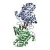
[English] 日本語
Yorodumi
- PDB-4f27: Crystal structures reveal the multi-ligand binding mechanism of t... -

+
Open data


ID or keywords:

Loading...

-
Basic information

Entry
Database: PDB / ID: 4f27
TitleCrystal structures reveal the multi-ligand binding mechanism of the Staphylococcus aureus ClfB
Components
  • Clumping factor B
  • peptide from Fibrinogen alpha chain
KeywordsCELL ADHESION/BLOOD CLOTTING / Dev-IgG fold / protein-peptide complex / fibronogen / cell surface / cell adhesion-blood clotting complex
Function / homology
Function and homology information


blood coagulation, common pathway / induction of bacterial agglutination / fibrinogen complex / Regulation of TLR by endogenous ligand / platelet alpha granule / blood coagulation, fibrin clot formation / positive regulation of heterotypic cell-cell adhesion / MyD88 deficiency (TLR2/4) / IRAK4 deficiency (TLR2/4) / extracellular matrix structural constituent ...blood coagulation, common pathway / induction of bacterial agglutination / fibrinogen complex / Regulation of TLR by endogenous ligand / platelet alpha granule / blood coagulation, fibrin clot formation / positive regulation of heterotypic cell-cell adhesion / MyD88 deficiency (TLR2/4) / IRAK4 deficiency (TLR2/4) / extracellular matrix structural constituent / MyD88:MAL(TIRAP) cascade initiated on plasma membrane / plasminogen activation / p130Cas linkage to MAPK signaling for integrins / positive regulation of peptide hormone secretion / positive regulation of exocytosis / GRB2:SOS provides linkage to MAPK signaling for Integrins / protein polymerization / Integrin cell surface interactions / Common Pathway of Fibrin Clot Formation / negative regulation of endothelial cell apoptotic process / negative regulation of extrinsic apoptotic signaling pathway via death domain receptors / fibrinolysis / positive regulation of vasoconstriction / positive regulation of substrate adhesion-dependent cell spreading / Integrin signaling / platelet alpha granule lumen / cell-matrix adhesion / positive regulation of protein secretion / Post-translational protein phosphorylation / Signaling by high-kinase activity BRAF mutants / MAP2K and MAPK activation / platelet aggregation / response to calcium ion / Regulation of Insulin-like Growth Factor (IGF) transport and uptake by Insulin-like Growth Factor Binding Proteins (IGFBPs) / Signaling by RAF1 mutants / Signaling by moderate kinase activity BRAF mutants / Paradoxical activation of RAF signaling by kinase inactive BRAF / Signaling downstream of RAS mutants / Signaling by BRAF and RAF1 fusions / Platelet degranulation / extracellular vesicle / ER-Phagosome pathway / protein-containing complex assembly / protein-macromolecule adaptor activity / : / blood microparticle / adaptive immune response / positive regulation of ERK1 and ERK2 cascade / cell adhesion / Amyloid fiber formation / endoplasmic reticulum lumen / external side of plasma membrane / signaling receptor binding / innate immune response / structural molecule activity / cell surface / endoplasmic reticulum / extracellular space / extracellular exosome / extracellular region / metal ion binding / plasma membrane
Similarity search - Function
: / Fibrinogen-binding domain 2 / C-terminus of bacterial fibrinogen-binding adhesin / Immunoglobulin-like - #1290 / SDR-like Ig domain / Bacterial Ig domain / Immunoglobulin-like - #1280 / Fibrinogen alpha C domain / Fibrinogen alpha C domain / Fibrogen-binding domain 1 ...: / Fibrinogen-binding domain 2 / C-terminus of bacterial fibrinogen-binding adhesin / Immunoglobulin-like - #1290 / SDR-like Ig domain / Bacterial Ig domain / Immunoglobulin-like - #1280 / Fibrinogen alpha C domain / Fibrinogen alpha C domain / Fibrogen-binding domain 1 / Fibrinogen, alpha/beta/gamma chain, coiled coil domain / Fibrinogen alpha/beta chain family / Fibrinogen alpha/beta chain family / Fibrinogen alpha chain / Fibrinogen, conserved site / Fibrinogen C-terminal domain signature. / Fibrinogen-related domains (FReDs) / Fibrinogen beta and gamma chains, C-terminal globular domain / Fibrinogen, alpha/beta/gamma chain, C-terminal globular, subdomain 1 / Fibrinogen, alpha/beta/gamma chain, C-terminal globular domain / Fibrinogen-like, C-terminal / Fibrinogen C-terminal domain profile. / Adhesion domain superfamily / YSIRK type signal peptide / YSIRK Gram-positive signal peptide / LPXTG cell wall anchor motif / Gram-positive cocci surface proteins LPxTG motif profile. / LPXTG cell wall anchor domain / Immunoglobulin-like / Sandwich / Mainly Beta
Similarity search - Domain/homology
Fibrinogen alpha chain / Clumping factor B
Similarity search - Component
Biological speciesStaphylococcus aureus (bacteria)
Homo sapiens (human)
MethodX-RAY DIFFRACTION / SYNCHROTRON / SAD / Resolution: 1.917 Å
AuthorsYang, M.J. / Xiang, H. / Wang, J.W. / Liu, B. / Chen, Y.G. / Liu, L. / Deng, X.M. / Feng, Y.
CitationJournal: Plos Pathog. / Year: 2012
Title: Crystal Structures Reveal the Multi-Ligand Binding Mechanism of Staphylococcus aureus ClfB
Authors: Xiang, H. / Feng, Y. / Wang, J.W. / Liu, B. / Chen, Y.G. / Liu, L. / Deng, X.M. / Yang, M.J.
History
DepositionMay 7, 2012Deposition site: RCSB / Processing site: PDBJ
Revision 1.0Aug 8, 2012Provider: repository / Type: Initial release
Revision 1.1Feb 4, 2015Group: Experimental preparation
Revision 1.2Oct 9, 2024Group: Data collection / Database references ...Data collection / Database references / Derived calculations / Structure summary
Category: chem_comp_atom / chem_comp_bond ...chem_comp_atom / chem_comp_bond / database_2 / pdbx_entry_details / pdbx_modification_feature / pdbx_struct_conn_angle / struct_conn / struct_ref_seq_dif / struct_site
Item: _database_2.pdbx_DOI / _database_2.pdbx_database_accession ..._database_2.pdbx_DOI / _database_2.pdbx_database_accession / _pdbx_struct_conn_angle.ptnr1_auth_comp_id / _pdbx_struct_conn_angle.ptnr1_auth_seq_id / _pdbx_struct_conn_angle.ptnr1_label_comp_id / _pdbx_struct_conn_angle.ptnr1_label_seq_id / _pdbx_struct_conn_angle.ptnr3_auth_comp_id / _pdbx_struct_conn_angle.ptnr3_auth_seq_id / _pdbx_struct_conn_angle.ptnr3_label_comp_id / _pdbx_struct_conn_angle.ptnr3_label_seq_id / _pdbx_struct_conn_angle.value / _struct_conn.pdbx_dist_value / _struct_conn.pdbx_leaving_atom_flag / _struct_conn.ptnr1_auth_comp_id / _struct_conn.ptnr1_auth_seq_id / _struct_conn.ptnr1_label_asym_id / _struct_conn.ptnr1_label_comp_id / _struct_conn.ptnr1_label_seq_id / _struct_conn.ptnr2_auth_seq_id / _struct_conn.ptnr2_label_asym_id / _struct_ref_seq_dif.details / _struct_site.pdbx_auth_asym_id / _struct_site.pdbx_auth_comp_id / _struct_site.pdbx_auth_seq_id

-
Structure visualization

Structure viewerMolecule:
MolmilJmol/JSmol

Downloads & links

-
Assembly

Deposited unit
A: Clumping factor B
Q: peptide from Fibrinogen alpha chain
hetero molecules


Theoretical massNumber of molelcules
Total (without water)41,2224
Polymers41,1732
Non-polymers492
Water2,090116
1


  • Idetical with deposited unit
  • defined by author&software
TypeNameSymmetry operationNumber
identity operation1_555x,y,z1
Buried area1830 Å2
ΔGint-20 kcal/mol
Surface area15080 Å2
MethodPISA
Unit cell
Length a, b, c (Å)70.983, 70.983, 174.908
Angle α, β, γ (deg.)90.00, 90.00, 120.00
Int Tables number152
Space group name H-MP3121

-
Components

#1: Protein Clumping factor B / Fibrinogen receptor B / Fibrinogen-binding protein B


Mass: 40063.094 Da / Num. of mol.: 1 / Fragment: UNP residues 197-542
Source method: isolated from a genetically manipulated source
Source: (gene. exp.) Staphylococcus aureus (bacteria) / Strain: MRSA252 / Gene: clfB, SAR2709 / Production host: Escherichia coli (E. coli) / References: UniProt: Q6GDH2
#2: Protein/peptide peptide from Fibrinogen alpha chain / Fibrinopeptide A / Fibrinogen alpha chain


Mass: 1110.051 Da / Num. of mol.: 1 / Source method: obtained synthetically / Details: This sequence occurs naturally in humans. / Source: (synth.) Homo sapiens (human) / References: UniProt: P02671
#3: Chemical ChemComp-MG / MAGNESIUM ION


Mass: 24.305 Da / Num. of mol.: 2 / Source method: obtained synthetically / Formula: Mg
#4: Water ChemComp-HOH / water


Mass: 18.015 Da / Num. of mol.: 116 / Source method: isolated from a natural source / Formula: H2O
Has protein modificationY

-
Experimental details

-
Experiment

ExperimentMethod: X-RAY DIFFRACTION / Number of used crystals: 1

-
Sample preparation

CrystalDensity Matthews: 3.09 Å3/Da / Density % sol: 60.19 %
Crystal growTemperature: 291 K / Method: vapor diffusion, hanging drop / pH: 5.6
Details: 0.1M sodium citrate tribasic dehydrate, 20% 2-propanol, 20% PEG 4000, pH 5.6, VAPOR DIFFUSION, HANGING DROP, temperature 291K

-
Data collection

DiffractionMean temperature: 100 K
Diffraction sourceSource: SYNCHROTRON / Site: SSRF / Beamline: BL17U / Wavelength: 0.979 Å
DetectorType: ADSC QUANTUM 315 / Detector: CCD / Date: Nov 1, 2009
RadiationMonochromator: Si 111 / Protocol: SINGLE WAVELENGTH / Monochromatic (M) / Laue (L): M / Scattering type: x-ray
Radiation wavelengthWavelength: 0.979 Å / Relative weight: 1
ReflectionResolution: 1.917→34.783 Å / Num. obs: 45741 / % possible obs: 100 % / Observed criterion σ(F): 0 / Observed criterion σ(I): 0 / Biso Wilson estimate: 40.38 Å2
Reflection shell
Resolution (Å)Diffraction-ID% possible all
1.9166-1.9684174
1.9684-2.0263188
2.0263-2.0917190
2.0917-2.1664193
2.1664-2.2532186
2.2532-2.3557188

-
Processing

Software
NameVersionClassification
HKL-2000data collection
SHELXSphasing
PHENIX(phenix.refine)refinement
HKL-2000data reduction
HKL-2000data scaling
RefinementMethod to determine structure: SAD / Resolution: 1.917→34.783 Å / Occupancy max: 1 / Occupancy min: 1 / SU ML: 0.2 / σ(F): 1.34 / Phase error: 24.98 / Stereochemistry target values: ML
RfactorNum. reflection% reflection
Rfree0.2259 1985 4.99 %
Rwork0.1891 --
obs0.1909 39740 99.12 %
Solvent computationShrinkage radii: 0.9 Å / VDW probe radii: 1.11 Å / Solvent model: FLAT BULK SOLVENT MODEL
Displacement parametersBiso mean: 51.1532 Å2
Baniso -1Baniso -2Baniso -3
1-4.7247 Å2-0 Å2-0 Å2
2--4.7247 Å20 Å2
3----9.4493 Å2
Refinement stepCycle: LAST / Resolution: 1.917→34.783 Å
ProteinNucleic acidLigandSolventTotal
Num. atoms2636 0 2 116 2754
Refine LS restraints
Refine-IDTypeDev idealNumber
X-RAY DIFFRACTIONf_bond_d0.0122683
X-RAY DIFFRACTIONf_angle_d1.3313637
X-RAY DIFFRACTIONf_dihedral_angle_d12.162964
X-RAY DIFFRACTIONf_chiral_restr0.099402
X-RAY DIFFRACTIONf_plane_restr0.006482
LS refinement shell

Refine-ID: X-RAY DIFFRACTION / Total num. of bins used: 14

Resolution (Å)Rfactor RfreeNum. reflection RfreeRfactor RworkNum. reflection RworkNum. reflection all% reflection obs (%)
1.9166-1.96450.31391370.292517265495
1.9645-2.01760.30511170.244126972814100
2.0176-2.0770.28051490.21926452794100
2.077-2.1440.21581430.210426632806100
2.144-2.22060.24881270.21962683281099
2.2206-2.30950.29371380.27292579271797
2.3095-2.41460.28671380.223327042842100
2.4146-2.54180.26961370.214727122849100
2.5418-2.7010.23291330.212626992832100
2.701-2.90950.24661600.217326962856100
2.9095-3.20210.28071500.207527372887100
3.2021-3.6650.21651750.191927072882100
3.665-4.61580.1931540.154927662920100
4.6158-34.78840.18311270.158729503077100
Refinement TLS params.Method: refined / Origin x: -35.1062 Å / Origin y: 0.3901 Å / Origin z: -12.9097 Å
111213212223313233
T0.5523 Å2-0.0902 Å2-0.0427 Å2-0.2466 Å20.0056 Å2--0.2754 Å2
L1.1842 °20.2751 °2-0.166 °2-1.4824 °2-0.4097 °2--1.432 °2
S0.0837 Å °-0.181 Å °0.0127 Å °-0.1206 Å °-0.1634 Å °-0.0135 Å °-0.0953 Å °0.0267 Å °0.0997 Å °
Refinement TLS groupSelection details: all

+
About Yorodumi

-
News

-
Feb 9, 2022. New format data for meta-information of EMDB entries

New format data for meta-information of EMDB entries

  • Version 3 of the EMDB header file is now the official format.
  • The previous official version 1.9 will be removed from the archive.

Related info.:EMDB header

External links:wwPDB to switch to version 3 of the EMDB data model

-
Aug 12, 2020. Covid-19 info

Covid-19 info

URL: https://pdbj.org/emnavi/covid19.php

New page: Covid-19 featured information page in EM Navigator.

Related info.:Covid-19 info / Mar 5, 2020. Novel coronavirus structure data

+
Mar 5, 2020. Novel coronavirus structure data

Novel coronavirus structure data

Related info.:Yorodumi Speices / Aug 12, 2020. Covid-19 info

External links:COVID-19 featured content - PDBj / Molecule of the Month (242):Coronavirus Proteases

+
Jan 31, 2019. EMDB accession codes are about to change! (news from PDBe EMDB page)

EMDB accession codes are about to change! (news from PDBe EMDB page)

  • The allocation of 4 digits for EMDB accession codes will soon come to an end. Whilst these codes will remain in use, new EMDB accession codes will include an additional digit and will expand incrementally as the available range of codes is exhausted. The current 4-digit format prefixed with “EMD-” (i.e. EMD-XXXX) will advance to a 5-digit format (i.e. EMD-XXXXX), and so on. It is currently estimated that the 4-digit codes will be depleted around Spring 2019, at which point the 5-digit format will come into force.
  • The EM Navigator/Yorodumi systems omit the EMD- prefix.

Related info.:Q: What is EMD? / ID/Accession-code notation in Yorodumi/EM Navigator

External links:EMDB Accession Codes are Changing Soon! / Contact to PDBj

+
Jul 12, 2017. Major update of PDB

Major update of PDB

  • wwPDB released updated PDB data conforming to the new PDBx/mmCIF dictionary.
  • This is a major update changing the version number from 4 to 5, and with Remediation, in which all the entries are updated.
  • In this update, many items about electron microscopy experimental information are reorganized (e.g. em_software).
  • Now, EM Navigator and Yorodumi are based on the updated data.

External links:wwPDB Remediation / Enriched Model Files Conforming to OneDep Data Standards Now Available in the PDB FTP Archive

-
Yorodumi

Thousand views of thousand structures

  • Yorodumi is a browser for structure data from EMDB, PDB, SASBDB, etc.
  • This page is also the successor to EM Navigator detail page, and also detail information page/front-end page for Omokage search.
  • The word "yorodu" (or yorozu) is an old Japanese word meaning "ten thousand". "mi" (miru) is to see.

Related info.:EMDB / PDB / SASBDB / Comparison of 3 databanks / Yorodumi Search / Aug 31, 2016. New EM Navigator & Yorodumi / Yorodumi Papers / Jmol/JSmol / Function and homology information / Changes in new EM Navigator and Yorodumi

Read more