Mass: 18.015 Da / Num. of mol.: 82 / Source method: isolated from a natural source / Formula: H2O
Has protein modification
Y
-
Experimental details
-
Experiment
Experiment
Method: X-RAY DIFFRACTION
-
Sample preparation
Crystal
Density Matthews: 2.74 Å3/Da / Density % sol: 55.06 % / Description: NONE
Crystal grow
Temperature: 298 K / Method: vapor diffusion, sitting drop Details: CRYSTALLIZATION TRIALS WERE PERFORMED AT 18 C BY THE SITTING DROP VAPOUR DIFFUSION METHOD. CRYSTALS GROWN BY MIXING 1 UL OF FC-SIAL (9 MG/ML IN PBS) WITH 3 UL OF PRECIPITANT SOLUTION ...Details: CRYSTALLIZATION TRIALS WERE PERFORMED AT 18 C BY THE SITTING DROP VAPOUR DIFFUSION METHOD. CRYSTALS GROWN BY MIXING 1 UL OF FC-SIAL (9 MG/ML IN PBS) WITH 3 UL OF PRECIPITANT SOLUTION CONSISTING OF 0.1M HEPES PH7, 16% PEG 6000, VAPOR DIFFUSION, TEMPERATURE 298K
Method to determine structure: OTHER Starting model: NONE Resolution: 2.3→20.1 Å / Cor.coef. Fo:Fc: 0.923 / Cor.coef. Fo:Fc free: 0.887 / SU B: 8.002 / SU ML: 0.194 / Cross valid method: THROUGHOUT / ESU R: 0.333 / ESU R Free: 0.255 / Stereochemistry target values: MAXIMUM LIKELIHOOD / Details: HYDROGENS HAVE BEEN ADDED IN THE RIDING POSITIONS.
Rfactor
Num. reflection
% reflection
Selection details
Rfree
0.28215
1300
5.1 %
RANDOM
Rwork
0.23398
-
-
-
obs
0.23647
24279
99.96 %
-
Solvent computation
Ion probe radii: 0.8 Å / Shrinkage radii: 0.8 Å / VDW probe radii: 1.2 Å / Solvent model: MASK
 Movie
Movie Controller

 Yorodumi
 Open data
 Basic information
 Components
 Keywords
 Function and homology information
 HOMO SAPIENS (human)
 X-RAY DIFFRACTION /
  Authors
 Citation
 Structure visualization
 Downloads & links
 Other downloads


 PDBj
PDBj


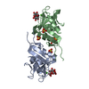
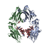
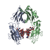
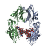
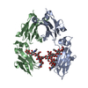
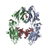
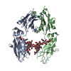
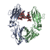
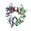
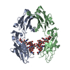
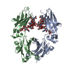
 Assembly
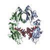
 Mass: 18.015 Da / Num. of mol.: 82 / Source method: isolated from a natural source / Formula: H2O
 Sample preparation
 / Beamline: ID23-1 / Type:
  Processing