 Movie
Movie Controller


+ Open data

- Basic information
| Entry | Database: PDB / ID: 1h3x | |||||||||
|---|---|---|---|---|---|---|---|---|---|---|
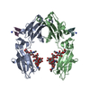
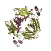
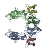
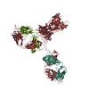
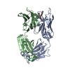
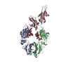
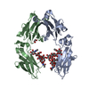
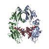
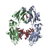
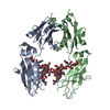
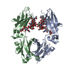
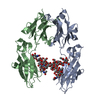
| Title | CRYSTAL STRUCTURE OF THE HUMAN IGG1 FC-FRAGMENT,GLYCOFORM (G0F)2 | |||||||||
 Components Components | IG GAMMA-1 CHAIN C REGION | |||||||||
 Keywords Keywords | IMMUNE SYSTEM / IMMUNOGLOBIN/FC-FRAGMENT / FC-FRAGMENT / GLYCOSYLATION / FCGR / ANTIBODY / EFFECTOR FUNCTIONS | |||||||||
| Function / homology |  Function and homology information Function and homology informationcomplement-dependent cytotoxicity / antibody-dependent cellular cytotoxicity / Fc-gamma receptor I complex binding / immunoglobulin complex, circulating / Classical antibody-mediated complement activation / immunoglobulin receptor binding / IgG immunoglobulin complex / Initial triggering of complement / FCGR activation / complement activation, classical pathway ...complement-dependent cytotoxicity / antibody-dependent cellular cytotoxicity / Fc-gamma receptor I complex binding / immunoglobulin complex, circulating / Classical antibody-mediated complement activation / immunoglobulin receptor binding / IgG immunoglobulin complex / Initial triggering of complement / FCGR activation / complement activation, classical pathway / Role of phospholipids in phagocytosis / antigen binding / FCGR3A-mediated IL10 synthesis / Regulation of Complement cascade / B cell receptor signaling pathway / FCGR3A-mediated phagocytosis / Regulation of actin dynamics for phagocytic cup formation / antibacterial humoral response / Interleukin-4 and Interleukin-13 signaling / blood microparticle / adaptive immune response / : / extracellular exosome / extracellular region / plasma membrane Similarity search - Function | |||||||||
| Biological species |  HOMO SAPIENS (human) HOMO SAPIENS (human) | |||||||||
| Method |  X-RAY DIFFRACTION / MOLECULAR REPLACEMENT / Resolution: 2.44 Å X-RAY DIFFRACTION / MOLECULAR REPLACEMENT / Resolution: 2.44 Å | |||||||||
 Authors Authors | Krapp, S. / Mimura, Y. / Jefferis, R. / Huber, R. / Sondermann, P. | |||||||||
 Citation Citation | Journal: J.Mol.Biol. / Year: 2003 Title: Structural Analysis of Human Igg-Fc Glycoforms Reveals a Correlation between Glycosylation and Structural Integrity Authors: Krapp, S. / Mimura, Y. / Jefferis, R. / Huber, R. / Sondermann, P. | |||||||||
| History |
| |||||||||
| Remark 700 | SHEET THE SHEET STRUCTURE OF THIS MOLECULE IS BIFURCATED. IN ORDER TO REPRESENT THIS FEATURE IN ... SHEET THE SHEET STRUCTURE OF THIS MOLECULE IS BIFURCATED. IN ORDER TO REPRESENT THIS FEATURE IN THE SHEET RECORDS BELOW, TWO SHEETS ARE DEFINED. |
- Structure visualization
| Structure viewer | Molecule: MolmilJmol/JSmol |
|---|
- Downloads & links
-Download
| PDBx/mmCIF format | 1h3x.cif.gz | 106 KB | Display | PDBx/mmCIF format |
|---|---|---|---|---|
| PDB format | pdb1h3x.ent.gz | 81.6 KB | Display | PDB format |
| PDBx/mmJSON format | 1h3x.json.gz | Tree view | PDBx/mmJSON format | |
| Others |  Other downloads Other downloads |
-Validation report
| Arichive directory | https://data.pdbj.org/pub/pdb/validation_reports/h3/1h3xftp://data.pdbj.org/pub/pdb/validation_reports/h3/1h3x | HTTPS FTP |
|---|
-Related structure data
| Related structure data |  1h3tC  1h3uC  1h3vC  1h3wC  1h3yC  1fc1S C: citing same article ( S: Starting model for refinement |
|---|---|
| Similar structure data |


-Links
 PDBj
PDBj


- Assembly
| Deposited unit | 
| ||||||||
|---|---|---|---|---|---|---|---|---|---|
| 1 |
| ||||||||
| Unit cell |
|
-Components
| #1: Protein | Mass: 25126.541 Da / Num. of mol.: 2 / Fragment: CH2, CH3, RESIDUES 225-447 / Source method: isolated from a natural source / Source: (natural) HOMO SAPIENS (human) / References: UniProt: P01857*PLUS#2: Polysaccharide | Source method: isolated from a genetically manipulated source #3: Water | ChemComp-HOH / |  Mass: 18.015 Da / Num. of mol.: 154 / Source method: isolated from a natural source / Formula: H2O Mass: 18.015 Da / Num. of mol.: 154 / Source method: isolated from a natural source / Formula: H2OHas protein modification | Y | Sequence details | THE PRIMARY SEQUENCE FOR THIS ENTRY IS VERY SIMILAR TO SWISSPROT ENTRY P01857 (GC1_HUMAN) WITH 96% ...THE PRIMARY SEQUENCE FOR THIS ENTRY IS VERY SIMILAR TO SWISSPROT ENTRY P01857 (GC1_HUMAN) WITH 96% SEQUENCE IDENTITY. | |
|---|
-Experimental details
-Experiment
| Experiment | Method: X-RAY DIFFRACTION / Number of used crystals: 1 |
|---|
- Sample preparation
| Crystal | Density Matthews: 2.75 Å3/Da / Density % sol: 55 % |
|---|---|
| Crystal grow | pH: 6.5 / Details: DIALYSIS AGAINST WATER, pH 6.50 |
-Data collection
| Diffraction | Mean temperature: 100 K |
|---|---|
| Diffraction source | Source: ROTATING ANODE / Type: RIGAKU RU200 / Wavelength: 1.5418 |
| Detector | Type: MARRESEARCH / Detector: IMAGE PLATE / Date: Aug 15, 2001 / Details: OS-MIRRORS |
| Radiation | Monochromator: GRAPHITE / Protocol: SINGLE WAVELENGTH / Monochromatic (M) / Laue (L): M / Scattering type: x-ray |
| Radiation wavelength | Wavelength: 1.5418 Å / Relative weight: 1 |
| Reflection | Resolution: 2.44→50 Å / Num. obs: 19885 / % possible obs: 93.6 % / Observed criterion σ(I): 2 / Redundancy: 3.2 % / Rmerge(I) obs: 0.074 |
| Reflection shell | Rmerge(I) obs: 0.41 / % possible all: 72.8 |
- Processing
| Software |
| ||||||||||||||||||||||||||||||||||||||||||||||||||||||||||||
|---|---|---|---|---|---|---|---|---|---|---|---|---|---|---|---|---|---|---|---|---|---|---|---|---|---|---|---|---|---|---|---|---|---|---|---|---|---|---|---|---|---|---|---|---|---|---|---|---|---|---|---|---|---|---|---|---|---|---|---|---|---|
| Refinement | Method to determine structure: MOLECULAR REPLACEMENT Starting model: PDB ENTRY 1FC1 Resolution: 2.44→50 Å / Cross valid method: THROUGHOUT / σ(F): 0
| ||||||||||||||||||||||||||||||||||||||||||||||||||||||||||||
| Refinement step | Cycle: LAST / Resolution: 2.44→50 Å
| ||||||||||||||||||||||||||||||||||||||||||||||||||||||||||||
| Refine LS restraints |
|
